| CAS NO: | 117570-53-3 |
| 规格: | ≥98% |
| 包装 | 价格(元) |
| 2mg | 电议 |
| 5mg | 电议 |
| 10mg | 电议 |
| 25mg | 电议 |
| 50mg | 电议 |
| 100mg | 电议 |
| 250mg | 电议 |
| Molecular Weight (MW) | 282.29 |
|---|---|
| Formula | C17H14O4 |
| CAS No. | 117570-53-3 |
| Storage | -20℃ for 3 years in powder form |
| -80℃ for 2 years in solvent | |
| Solubility (In vitro) | DMSO: 7 mg/mL (24.8 mM) |
| Water: <1 mg/mL | |
| Ethanol: <1 mg/mL | |
| Solubility (In vivo) | 30% PEG400+0.5% Tween80+5% propylene glycol: 30 mg/mL |
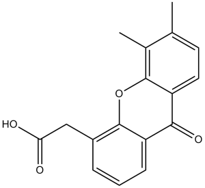
| Synonyms | NSC 640488, NSC-640488, NSC640488, ASA-404, Vadimezan; ASA404; ASA 404; AS1404; AS 1404; AS1404; DMXAA zChemical Name: (5,6-Dimethyl-9-oxo-9H-xanthen-4-yl)-acetic acid SMILES Code: O=C(O)CC1=CC=CC(C2=O)=C1OC3=C2C=CC(C)=C3C |
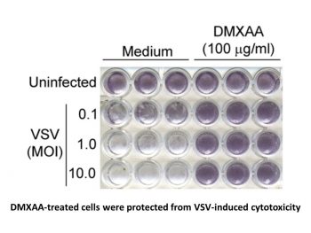
| In Vitro | In vitro activity: In DLD-1 human colon carcinoma cells, DMXAA inhibits DT-diaphorase activity without significant effects on the activity of cytochrome b5 reductase and cytochrome P450 reductase. Combination of menadione and DMXAA leads to an increase in the antiproliferative activity of DLD-1 cells. DMXAA, as an antiviral agent, inhibits VSV-induced cytotoxicity and influenza virus replication in RAW 264.7 macrophages. A recent study shows that DMXAA has non-immune-mediated inhibitory effects against several kinase members of VEGFR (vascular endothelial growth factor receptor), such as VEGFR2 signalling in human umbilical vein endothelial cells. Kinase Assay: Purified DT-diaphorase enzyme activity is assayed by measuring the reduction of cytochrome c at 550 nm on a Beckman DU 650 spectrophotometer. Each assay contains cytochrome c (70 μM), NADH (variable concentrations), purified DT-diaphorase (0.032 μg), and menadione (variable concentrations) in a final volume of 1 mL Tris–HCl buffer (50 mM, pH 7.4) containing 0.14% BSA. The reaction is started by the addition of NADH. Rates of reduction are calculated over the initial part of the reaction curve (30 seconds), and results are expressed in terms of μmol cytochrome c reduced/min/mg protein using a molar extinction coefficient of 21.1 mM–1 cm–1 for reduced cytochrome c. Enzyme assays are carried out at room temperature and all reactions are performed in triplicate. Inhibition of purified DT-diaphorase activity is performed by the inclusion of DMXAA (at various concentrations) in the reaction, and inhibition characteristics are determined by varying the concentration of NADH (constant menadione) or menadione (constant NADH) at several concentrations of inhibitor. Ki values are obtained by plotting 1/V against. The activity of DT-diaphorase in DLD-1 cells is determined by measuring the dicumarol-sensitive reduction of DCPIP at 600 nm. Briefly, DLD-1 cells in mid-exponential growth are harvested by scraping into ice-cold buffer (Tris–HCl, 25 mM, pH 7.4 and 250 mM sucrose) and sonicated on ice. Enzyme assay conditions are 2 mM NADH, 40 μM DCPIP, 20 μL of dicumarol (when required) in a final volume of 1 mL Tris–HCl (25 mM, pH 7.4) containing BSA (0.7 mg/mL). Results are expressed as the dicumarol-sensitive reduction of DCPIP using a molar extinction coefficient of 21 mM–1 cm–1. Protein levels are determined using the Bradford assay. Cell Assay: DLD-1 human colon carcinoma and H460 human non-small cell lung carcinoma cells are routinely maintained as monolayer cultures in RPMI 1640 culture medium supplemented with foetal calf serum (10%), sodium pyruvate (2 mM), penicillin/streptomycin (50 IU mL–1/50 μg mL-1) and l-glutamine (2 mM). Chemosensitivity is assessed using the MTT assay and all assays are performed under aerobic conditions. Briefly, cells are plated into each well of a 96-well culture plate and incubated overnight in an atmosphere containing 5% CO2. Culture medium is completely removed from each well and replaced with medium containing a range of drug concentrations. In the case of menadione alone, the duration of drug exposure is 1 hour, after which the cells are washed twice with Hanks' balanced salt solution prior to the addition of 200 μL fresh RPMI 1640 medium to each well of the plate. In the case of DMXAA alone, the duration of drug exposure is 3 hours. Following a four-day incubation, cell survival is determined using the MTT assay. For combinations of DMXAA with menadione, cells are initially set up and a non-toxic (>95% cell survival) concentration of DMXAA is selected. Cells are then initially exposed to DMXAA (2 mM) for a period of 2 hours, following which the medium is removed and replaced with medium containing the inhibitor (DMXAA at a constant concentration of 2 mM) and menadione (at a range of drug concentrations). Following a further 7-hour incubation, cells are washed twice with Hanks' balanced salt solution prior to the addition of growth medium. |
|---|---|
| In Vivo | DMXAA treatment significantly protects C57BL/6J mice infected i.n. with 200 p.f.u. mouse-adapted H1N1 influenza PR8 virus with 60% survival, while the control group only exhibited 20% survival. DMXAA significantly delays tumor growth induced by chemical carcinogen, increases the time to tumor doubling and increases time from treatment to euthanasia. After the treatment of DMXAA, median tumor doubling time, median tumour tripling time and median time from treatment to euthanasia in tumor-bearing animals are increased by approximately 4.4-, 1.8- and 2.7-fold, respectively. |
| Animal model | Chemical carcinogen (NMU) is injected into female Wistar rats. |
| Formulation & Dosage | Dissolved in in 5% sodium bicarbonate; 300mg/kg; i.p. injection |
| References | J Leukoc Biol. 2011 Mar;89(3):351-7; Biochem Pharmacol. 1999 Jul 15;58(2):303-10. |
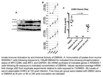
DMXAA inhibits influenza A/Wuhan and Tamiflu(R)-resistant influenza A/Br replication in vitro. J Leukoc Biol. 2011 Mar;89(3):351-7. |
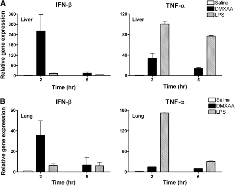
Differential induction of IFN-β and TNF-α mRNA expression by DMXAA and LPS in vivo. J Leukoc Biol. 2011 Mar;89(3):351-7. |
IFN-β-dependent, DMXAA-mediated protection of mice against influenza-induced lethality. J Leukoc Biol. 2011 Mar;89(3):351-7. |
 |  |  |
 m.cnreagent.com