| CAS NO: | 1020172-07-9 |
| 规格: | ≥98% |
| 包装 | 价格(元) |
| 5mg | 电议 |
| 10mg | 电议 |
| 25mg | 电议 |
| 50mg | 电议 |
| 100mg | 电议 |
| 250mg | 电议 |
| 500mg | 电议 |
| Molecular Weight (MW) | 553.59 |
|---|---|
| Formula | C30H28FN7O3 |
| CAS No. | 1020172-07-9 |
| Storage | -20℃ for 3 years in powder form |
| -80℃ for 2 years in solvent | |
| Solubility (In vitro) | DMSO: 111 mg/mL (200.5 mM) |
| Water:<1 mg/mL | |
| Ethanol: 16 mg/mL (28.9 mM) | |
| Solubility (In vivo) | 0.5% CMC+0.25% Tween 80: 16 mg/mL |
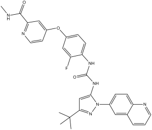
| Synonyms | Synonym: DCC-2036; DCC 2036; DCC2036; Rebastinib. Chemical Name: N-[3-tert-Butyl-1-(quinolin-6-yl)-1H-pyrazol-5-yl]-N'-[2-fluoro-4-[(2-(methylcarbamoyl)pyridin-4-yl)oxy]phenyl]urea InChi Key: WVXNSAVVKYZVOE-UHFFFAOYSA-N InChi Code: InChI=1S/C30H28FN7O3/c1-30(2,3)26-17-27(38(37-26)19-7-9-23-18(14-19)6-5-12-33-23)36-29(40)35-24-10-8-20(15-22(24)31)41-21-11-13-34-25(16-21)28(39)32-4/h5-17H,1-4H3,(H,32,39)(H2,35,36,40) SMILES Code: O=C(NC1=CC=C(OC2=CC(C(NC)=O)=NC=C2)C=C1F)NC3=CC(C(C)(C)C)=NN3C4=CC=C5N=CC=CC5=C4 |
| In Vitro | In vitro activity: DCC-2036 shows the potent inhibitory activities against purified native Abl1 in unphosphorylated (u-Abl1native) and phosphorylated (p-Abl1native) forms, unphosphorylated and phosphorylated gatekeeper mutant Abl1T315I, and the activation loop mutant Abl1H396P in a non-ATP-competitive manner with IC50 of 0.8 nM, 2 nM, 1.4 nM, 5 nM, and 4 nM, respectively. Moreover, DCC-2036 also inhibits the Src family kinases Src, LYN, FGR, and HCK, and the receptor TKs KDR, FLT3, and TIE2 with IC50 of 34 nM, 29 nM, 38 nM, 40 nM, 4 nM, 2 nM and 6 nM, respectively. DCC-2036 shows the anti-proliferative activities against Ba/F3 cells expressing native or mutant Bcr-Abl1 with IC50 ranging from 2 nM to 150 nM. In addition, DCC-2036 also inhibits proliferation of the Ph+ cell line K562 (IC50 5.5 nM), and induces apoptosis in both Bcr-Abl1-expressing Ba/F3 and K562 cells potently. A recent study shows that DCC-2036 shows the selectivity for growth inhibition of Bcr-Abl-positive cells by its marked inhibition of CML cell lines compared to non-CML leukemia lines. Kinase Assay: Activity of u-Abl1native is determined by following the production of ADP from the kinase reaction through coupling with the pyruvate kinase/lactate dehydrogenase system. In this assay, the oxidation of NADH (measured as a decreased A340nm) is continuously monitored spectrophotometrically. The final reaction mixture (100 μL, in a 384-well Corning plate) is prepared as follows: An Abl1 kinase/coupled assay components mixture is prepared containing u-Abl1 kinase (1 nM), Abltide (EAIYAAPFAKKK, 0.2 mM), MgCl2 (9 mM), pyruvate kinase (~ 4 units), lactate dehydrogenase (~ 0.7 units), phosphoenol pyruvate (1 mM), and NADH (0.28 mM) in 90 mM Tris containing 0.1 % octyl-glucoside and 1 % DMSO, pH 7.5. Separately, an inhibitor mixture is prepared containing DCC-2036 serially diluted 3-fold in DMSO followed by dilution into buffer composed of 180 mM Tris, pH 7.5, containing MgCl2 (18 mM) and 0.2 % octyl-glucoside. Fifty μL of the inhibitor mixture is mixed with 50 μL of the above Abl1 kinase/coupled assay components mixture, which is then incubated at 30 °C for 2 hours before 2 μL of 25 mM ATP (500 μM, final) is added to start the reaction. The reaction is recorded every 2 minutes for 2.5 hours at 30 °C on a Polarstar Optima or Synergy2 plate reader. Reaction rate (slope) is calculated using the 1 to 2 hour time frame with reader's software. Percent inhibition is obtained by comparison of reaction rate with that of a DMSO control. IC50 values are calculated from a series of percent inhibition values determined at a range of inhibitor concentrations using GraphPad Prism. The kinase assay for Abl1T315I, p-Abl1native or Abl1H396P is assayed the same as above except that 2.2 nM Abl1T315I, 1 nM p-Abl1 native or 1.3 nM Abl1H396P is used. The above assay format is also used for kinases other than Abl1 with the exception of TIE2, for which a fluorescence polarization/Transcreener format is used. The assay conditions are the same as described above except that PolyE4Y (final 1 mg/mL) is used as the substrate and one hour preincubation is used. Cell Assay: Ba/F3 cells or primary Ph+ leukemia cells are plated in triplicate in 96-well plates containing test compounds. After 72 hours, viable cells are quantified by Resazurin or MTT assay. Cells (Ba/F3 cells and primary Ph+ leukemia cells) are diluted in medium to be added to each well of a 96-well tissue culture-treated plate. All cells are incubated overnight and maintained in a humidified atmosphere at 37 °C and 5% CO2. Cells are treated the following day. Serum-free medium is used during treatment with DCC-2036. MTT is used to assess the viability of cells following treatment. Aliquots of 20 mL of stock MTT solution are added to each well containing 200 mL of medium (10% final solution) and incubated with the cells for 2 hours. Following incubation the medium is removed and 200 mL of dimethylsulfoxide added to solubilize the formazan crystals. The absorbance is read on the plate reader at 550 and 690 nm. A subtraction analysis of the dual wavelength is performed (D550 to D690) to increase accuracy of the measurement. |
|---|---|
| In Vivo | In a mouse allograft model bearing Ba/F3-Bcr-Abl1T315I leukemia cells, DCC-2036 treatment by oral gavage at 100 mg/kg once daily effectively inhibits Bcr-Abl1 signaling and significantly prolongs mouse survival. |
| Animal model | Ba/F3 cells transformed to interleukin-3 independence by transduction with either Bcr-Abl1native or Bcr-Abl1T315I retrovirus are injected intravenously into syngeneic Balb/c mice. |
| Formulation & Dosage | Dissolved in 0.5% CMC/1% Tween-80; ≤100 mg/kg; p.o. |
| References | Cancer Cell. 2011 Apr 12;19(4):556-68; Cancer Res. 2011 May 1;71(9):3189-95. |
 m.cnreagent.com