| CAS NO: | 172889-26-8 |
| 规格: | ≥98% |
| 包装 | 价格(元) |
| 10mg | 电议 |
| 25mg | 电议 |
| 50mg | 电议 |
| 100mg | 电议 |
| 250mg | 电议 |
| 500mg | 电议 |
| 1g | 电议 |
| Molecular Weight (MW) | 281.36 |
|---|---|
| Formula | C16H19N5 |
| CAS No. | 172889-26-8 |
| Storage | -20℃ for 3 years in powder form |
| -80℃ for 2 years in solvent | |
| Solubility (In vitro) | DMSO: 4 mg/mL (14.2 mM) |
| Water: <1 mg/mL | |
| Ethanol: <1 mg/mL | |
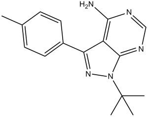
| SMILES | NC1=C2C(N(C(C)(C)C)N=C2C3=CC=C(C)C=C3)=NC=N1 |
| Synonyms | AGL 1872; EI 275; PP 1; AGL-1872; EI-275; PP-1; AGL1872; EI275; PP1 |
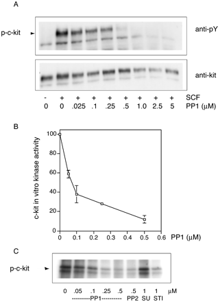
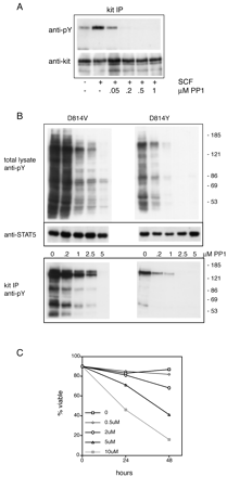
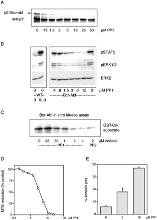
| In Vitro | In vitro activity: PP1 is a nano-molar inhibitor of Lck and FynT, inhibits anti-CD3-induced protein-tyrosine kinase activity in T cells (IC50, 0.5 μM), demonstrates selectivity for Lck and FynT over ZAP-70, and preferentially inhibits T cell receptor-dependent anti-CD3-induced T cell proliferation (IC50, 0. 5 μM) over non-T cell receptor-dependent phorbol 12-myristate 13-acetate/interleu-kin-2 (IL-2)-induced T cell proliferation. PP1 (1 μM) selectively inhibits the induction of the IL-2 gene, but not the granulocyte-macrophage colony-stimulating factor or IL-2 receptor genes. PP1 also inhibits Src (IC50, 170 nM) and Hck (IC50, 20 nM). PP1 is 50–100-fold less active in the inhibition of A-431 epidermal growth factor receptor autophosphorylation (IC50, 0.25 μM). PP1 also inhibits Kit and Bcr-Abl tyrosine kinases with IC50 of ~75 nM and 1 μM, respectively. PP1 completely abrogates the proliferation of M07e cells in response to SCF with IC50 of 0.5–1 μM. PP1 (1 μM) inhibits SCF-induced c-Kit autophosphorylation in intact cells and blocks the activation of mitogen-activated protein kinase and Akt. PP1 inhibits the activity of mutant constitutively active forms of c-Kit (D814V and D814Y) found in mast cell disorders, and triggers apoptosis in the rat basophilic leukemia cell line RBL-2H3 that expresses mutant c-Kit. PP1 reduces the constitutive activation of signal transducer and activators of transcription 5 and mitogen-activated protein kinase and triggeres apoptosis in FDCP1 cells expressing Bcr-Abl. Kinase Assay: Protein A-Sepharose beads (prepared as a 50% (w/v) suspension) are added to the antibody/lysate mixture at 250 μL/mL and allowed to incubate for 30 min at 4°C. The beads are then washed twice in 1 mL of lysis buffer and twice in 1 mL of kinase buffer (25 mM HEPES, 3 mM MnCl2, 5 mM MgCl2, and 100 μM sodium orthovanadate) and resuspended to 50% (w/v) in kinase buffer. Twenty-five microliters of the bead suspension is added to each well of the enolase-coated 96-well high protein binding plate together with an appropriate concentration of compound and [γ-32P]ATP (25 μL/well of a 200 μCi/mL solution in kinase buffer). After incubation for 20 min at 20°C, 60 μL of boiling 2× solubilization buffer containing 10 mM ATP is added to the assay wells to terminate the reactions. Thirty microliters of the samples is removed from the wells, boiled for 5 min, and run on a 7.5% SDS-polyacrylamide gel. The gels are subsequently dried and exposed to Kodak X-AR film. For quantitation, films are scanned using a Molecular Dynamics laser scanner, and the optical density of the major substrate band, enolase p46, is determined. Concentrations of compound that causes 50% inhibition of enolase phosphorylation (IC50) are determined from a plot of the density versus concentration of compound. In companion experiments for measuring the activity of compounds against Lck, the assay plate is washed with two wash cycles on a Skatron harvester using 50 mM EDTA, 1 mM ATP. Scintillation fluid (100 μL) is then added to the wells, and P incorporation is measured using a Pharmacia Biotech micro-β-counter. Concentrations of compound that causes 50% inhibition of enzyme activity (IC50) are determined from a plot of the percent inhibition of enzyme activity versus concentration of compound Cell Assay: Inhibition of anti-CD3-stimulated tyrosine phosphorylation in purified human peripheral blood T cells is measured as follows. All incubations are carried out at 37°C in an Eppendorf Thermomixer 5436 at a mixing setting of 11. Cells (1×10⁶ in 100 μL of RPMI 1640 medium) are incubated for 15 min with drug prior to a 6-min incubation with 1 μg of anti-CD3/mL (anti-leu4, 100 μg/mL). The final volume of the reaction is 115 μL. Reactions are terminated by the addition of 57.5 μL of 3× solubilization buffer incubated at 100°C prior to its addition. Samples are mixed, boiled for 5 min, and stored at -70°C. Western blots of these cell lysates, run on 10% SDS-polyacrylamide gels, are probed with a polyclonal anti-phosphotyrosine antibody, and immune complexes are detected with I-labeled protein A (ICN). For quantitation, films are scanned using a Molecular Dynamics laser scanner, and the optical densities of the major substrate band, p70, are quantitated in the presence of anti-CD3 (in the presence and absence of drug). Percent inhibition is calculated as follows: (1-(p70 optical density units in presence of drug/p70 units in absence of drug))×100. IC50 equals the concentration of compound at which 50% inhibition is measured. |
|---|---|
| In Vivo | Under in vivo conditions PP1 was suggested to suppress tyrosine phosphorylation and proliferation in T cells stimulated with anti-CD3 and mitogen. Studies using mice tumor model also showed that PP1 upregulated the expression of the IL-2 gene rather than the granulocyte macrophage colony-stimulating factor or the IL-2 receptor genes. Based on these, PP1 could be adopted as a useful agent to investigate the role of Lck and Fyn T cell activation. |
| Animal model | |
| Formulation & Dosage | |
| References | J Biol Chem. 1996 Jan 12;271(2):695-701; J Biol Chem. 2003 Feb 14;278(7):4847-53. |
|  |
PP1 inhibits c-Kit autophosphorylation in intact cells and in vitro kinase assays. J Biol Chem.2003 Feb 14;278(7):4847-53. |
Effects of PP1 , SU6656, and STI571 on c-Kit activation. J Biol Chem. 2003 Feb 14;278(7):4847-53. |
Effects of PP1 on the phosphorylation and activity of wild-type and constitutively active mutant forms of c-Kit. J Biol Chem. 2003 Feb 14;278(7):4847-53. |
PP1 blocks constitutive activation of Bcr-Abl and causes rapid apoptosis in Bcr-Abl/FDCP1 cells. J Biol Chem. 2003 Feb 14;278(7):4847-53. |
 m.cnreagent.com