| CAS NO: | 183321-74-6 |
| 规格: | ≥98% |
| 包装 | 价格(元) |
| 50mg | 电议 |
| 100mg | 电议 |
| 250mg | 电议 |
| 500mg | 电议 |
| 1g | 电议 |
| 5g | 电议 |
| 10g | 电议 |
| 25g | 电议 |
| Molecular Weight (MW) | 393.44 |
|---|---|
| Formula | C22H23N3O4 |
| CAS No. | 183321-74-6 (free base); |
| Storage | -20℃ for 3 years in powder form |
| -80℃ for 2 years in solvent | |
| Solubility (In vitro) | DMSO: 78 mg/mL (198.3 mM) |
| Water: <1 mg/mL | |
| Ethanol: 15 mg/mL (38.1 mM) | |
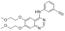
| SMILES Code | COCCOC1=CC2=NC=NC(NC3=CC=CC(C#C)=C3)=C2C=C1OCCOC |
| Synonyms | Erlotinib free base; NSC718781; NSC718781; CP358774; OSI-774; OSI 774; NSC 718781; CP-358,774; CP-358774; OSI774 Chemical Name: N-(3-Ethynylphenyl)-6,7-bis(2-methoxyethoxy)-4-quinazolinamine SMILES Code: COCCOC1=CC2=NC=NC(NC3=CC=CC(C#C)=C3)=C2C=C1OCCOC |
| In Vitro | In vitro activity: Erlotinib potently inhibits EGFR activation in intact cells including HNS human head and neck tumor cells (IC50 20nM), DiFi humancolon cancer cells andMDA MB-468 human breast cancer cells. Erlotinib (1 μM) induces apoptosis in DiFi humancolon cancer cells. Erlotinib inhibits growth of a panel of NSCLC cell lines including A549, H322, H3255, H358 H661, H1650, H1975, H1299, H596 with IC50 ranging from 29 nM to>20 μM. Erlotinib (2 μM) significantly inhibits growth of AsPC-1 and BxPC-3 pancreatic cells. The effects of Erlotinib in combination with gemcitabine are considered additive in KRAS-mutated pancreatic cancer cells. Ten micromolar of Erlotinib inhibits EGFR phospho-rylation at the Y845 (Src-dependent phosphorylation) and Y1068 (auto-phosphorylation) sites. Combination with Erlotinib could down-modulate rapamycin-stimulated Akt activity and produces a synergistic effect on cell growth inhibition. Kinase Assay: 96-well plates are coated by incubation overnight at 37 °C with 100 μL per well of 0.25 mg/mL PGT in PBS. Excess PGT is removed by aspiration, and the plate is washed 3 times with washing buffer (0.1% Tween 20 in PBS). The kinase reaction is performed in 50 μL of 50 mM HEPES (pH 7.3), containing 125 mM sodium chloride, 24 mM magnesium chloride, 0.1 mM sodium orthovanadate, 20 μM ATP, 1.6 μg/mL EGF, and 15 ng of EGFR, affinity purified from A431 cell membranes. Erlotinib in DMSO is added to give a final DMSO concentration of 2.5%. Phosphorylation is initiated by addition of ATP and proceeded for 8 minutes at room temperature, with constant shaking. The kinase reaction is terminated by aspiration of the reaction mixture and is washed 4 times with washing buffer. Phosphorylated PGT is measured by 25 minutes of incubation with 50 μL per well HRP-conjugated PY54 antiphosphotyrosine antibody, diluted to 0.2 μg/mL in blocking buffer (3% BSA and 0.05% Tween 20 in PBS). Antibody is removed by aspiration, and the plate is washed 4 times with washing buffer. The colonmetric signal is developed by addition of TMB Microwell Peroxidase Substrate, 50μL per well, and stopped by the addition of 0.09 M sulfuric acid, 50 μL per well. Phosphotyrosine is estimated by measurement of absorbance at 450 nm. The signal for controls is typically 0.6-1.2 absorbance units, with essentially no back ground in wells without AlP, EGFR, or PGT and is proportional to the time of incubation for 10 minutes. Cell Assay: Exponentially growing cells (A549, H322, H3255, H358 H661, H1650, H1975, H1299, H596 cells) are seeded in 96-well plastic plates and exposed to serial dilutions of erlotinib, pemetrexed, or the combination at a constant concentration ratio of 4:1 in triplicates for 72 h. Cell viability is assayed by cell count and the 3-(4,5-dimethylthiazol-2-yl)-2,5-diphenyltetrazolium bromide assay. Growth inhibition is expressed as the percentage of surviving cells in drug-treated versus PBS-treated control cells (which is considered as 100% viability). The IC50 value is the concentration resulting in 50% cell growth inhibition by a 72-h exposure to drug(s) compared with untreated control cells and is calculated by the CalcuSyn software. |
|---|---|
| In Vivo | At doses of 100 mg/kg, Erlotinib HCl completely prevents EGF-induced autophosphorylation of EGFR in human HN5 tumors growing as xenografts in athymic mice and of the hepatic EGFR of the treated mice. Erlotinib (100 mg/Kg) inhibits H460a and A549 tumor models with 71 and 93% inhibition rate. |
| Animal model | Male 5-week-old BALB-nu/nu with HPAC cells |
| Formulation & Dosage | Dissolved in 6% Captisol; 50 mg/kg; Oral gavage |
| References | Cancer Res. 1997 Nov 1;57(21):4838-48; Anticancer Drugs. 2004 Jun;15(5):503-12; Mol Cancer Ther. 2008 Jun;7(6):1708-19. |
 m.cnreagent.com