| CAS NO: | 252916-29-3 |
| 规格: | ≥98% |
| 包装 | 价格(元) |
| 5mg | 电议 |
| 10mg | 电议 |
| 25mg | 电议 |
| 50mg | 电议 |
| 100mg | 电议 |
| 250mg | 电议 |
| Molecular Weight (MW) | 310.35 |
|---|---|
| Formula | C18H18N2O3 |
| CAS No. | 252916-29-3 |
| Storage | -20℃ for 3 years in powder form |
| -80℃ for 2 years in solvent | |
| Solubility (In vitro) | DMSO: 62 mg/mL (199.77 mM) |
| Water: <1 mg/mL | |
| Ethanol:<1 mg/mL | |
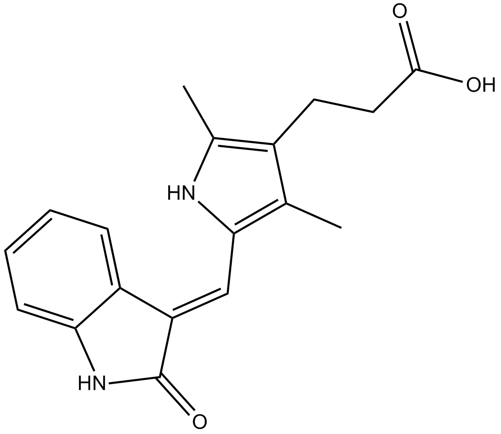
| SMILES | O=C(O)CCC1=C(C)NC(/C=C2C(NC3=C\2C=CC=C3)=O)=C1C |
| Synonyms | NSC 702827; NSC-702827; NSC702827; TSU68; TSU-68; SU-6668; SU 6668; SU6668; NSC702827; TSU 68; Chemical Name: (Z)-3-(2,4-dimethyl-5-((2-oxoindolin-3-ylidene)methyl)-1H-pyrrol-3-yl)propanoic acid Exact Mass: 482.19541 |
| In Vitro | In vitro activity: TSU-68 is a competitive inhibitor, with regard to ATP, to Flk-1/KDR trans-phosphorylation, FGFR1 trans-phosphorylation, and PDGFRβ kinases autophosphorylation. TSU-68 (0.03-10 μM) inhibits tyrosine phosphorylation of KDR in VEGF stimulated HUVECs. TSU-68 also inhibits PDGF-stimulated PDGFRβ tyrosine phosphorylation in NIH-3T3 cells overexpressing PDGFRβ at a minimum concentration of 0.03-0.1 μM. TSU-68 inhibits acidic FGF-induced phosphorylation of the FGFR1 substrate 2 at 10 μM and higher. However, TSU-68 (up to 100 μM) has no effect on EGF-stimulated EGFR tyrosine phosphorylation in NIH-3T3 cells overexpressing EGFR. TSU-68 inhibits VEGF-driven and FGF-driven mitogenesis of HUVECs with mean IC50 of 0.34 μM and 9.6 μM, respectively. In human myeloid leukemia MO7E cells, TSU-68 inhibits the tyrosine autophosphorylation of stem cell factor (SCF) receptor, c-kit, with IC50 of 0.1-1 μM, as well as ERK1/2 phosphorylation, a signaling event downstream of c-kit activation. TSU-68 also inhibits SCF-induced proliferation of MO7E cells with IC50 of 0.29 μM, and induces apoptosis. Kinase Assay: Tyrosine kinase assays to quantitate the trans-phosphorylation activity of Flk-1 and FGFR1 are performed in 96-well microtiter plates precoated (20 μg/well in PBS; incubated overnight at 4 °C) with the peptide substrate poly-Glu,Tyr (4:1). Excess protein binding sites are blocked with 1-5% (w/v) BSA in PBS. Purified GST-FGFR1 (kinase domain) or GST-Flk-1 (cytoplasmic domain) fusion proteins are then added to the microtiter wells in 2 × concentration kinase dilution buffer consisting of 100 mM HEPES, 50 mM NaCl, 40 μM NaVO4, and 0.02% (w/v) BSA. The final enzyme concentration for GST-Flk-1 and GST-FGFR1 is 50 ng/mL. SU6668 is dissolved in DMSO at 100× the final required concentration and diluted 1:25 in H2O. Twenty-five μL of diluted SU6668 are subsequently added to each reaction well. The kinase reaction is initiated by the addition of different concentrations of ATP in a solution of MnCl2 so that the final ATP concentrations spanned the Km for the enzyme, and the final concentration of MnCl2 is 10 mM. The plates are incubated for 5-15 min at room temperature before stopping the reaction with the addition of EDTA. The plates are then washed three times with TBST. Rabbit polyclonal antiphosphotyrosine antisera are added to the wells at a 1: 10000 dilution in TBST containing 0.5% (w/v) BSA, 0.025% (w/v) nonfat dry milk, and 100 μM NaVO4 and incubated for 1 hour at 37 °C. The plates are then washed three times with TBST, followed by the addition of goat anti-rabbit antisera conjugated with HRP. The plates are incubated for 1 hour at 37 °C and then washed three times with TBST. Cell Assay: Cells (HUVECs, and NIH-3T3 cells overexpressing PDGFRβ or EGFR) are seeded (3 × 10⁵ cells/35-mm well) in DMEM containing 10% (v/v) FBS and grow to confluence and then quiesced in DMEM containing 0.1% serum for 2 hours before drug treatment. HUVECs (seeded at 2 × 10⁶ cells/10-cm plate) are grown to confluence in endothelial cell growth media and then quiesced in endothelial cell basal media containing 0.5% FBS for 24 hours before drug treatment. All cell lines are incubated with SU6668 for 1 hour before ligand stimulation (100 ng/mL) for 10 min. |
|---|---|
| In Vivo | TSU-68 (75-200 mg/kg) induces tumor growth inhibition against a broad range of tumor types in xenograft models in athymic mice, including A375, Colo205, H460, Calu-6, C6, SF763T, and SKOV3TP5 cells. TSU-68 (75 mg/kg) also suppresses tumor angiogenesis of C6 glioma xenografts. In a tumor model of HT29 human colon carcinoma, TSU-68 (200 mg/kg) decreases the average vessel permeability and average fractional plasma volume in the tumor rim and core. TSU-68 promotes abnormal stromal development at the periphery of carcinomas. In a rabbit VX2 liver tumor model, TSU-68 (200 mg/kg) augments the effect of chemotherapeutic infusion. |
| Animal model | Female, BALB/c, nu/nu mouse xenograft models of A375, Colo205, H460, Calu-6, C6, SF763T, and SKOV3TP5 tumor cells |
| Formulation & Dosage | Dissolved in DMSO; 75-200 mg/kg; p.o. or i.p. injection |
| References | Cancer Res. 2000 Aug 1;60(15):4152-60; Clin Cancer Res. 2004 Jan 15;10(2):739-50; Cardiovasc Intervent Radiol. 2012 Feb;35(1):168-75. |
Efficacy of SU6668 on s.c. A431 xenograft growth in athymic mice. Cancer Res. 2000 Aug 1;60(15):4152-60. |
Effect of SU6668 on tumor xenograft angiogenesis. Cancer Res. 2000 Aug 1;60(15):4152-60. |
Efficacy of SU6668 against established A431 s.c. xenografts in athymic mice. A, SU6668 regresses established tumors in athymic mice. Cancer Res. 2000 Aug 1;60(15):4152-60. |
A, HUVECs; B, NIH-3T3 cells overexpressing PDGFRβ; C, NIH-3T3 cells; D, NIH-3T3 cells overexpressing EGFR. Cancer Res. 2000 Aug 1;60(15):4152-60. |
Inhibition of endothelial cell proliferation stimulated by either VEGF or FGF. Cancer Res. 2000 Aug 1;60(15):4152-60. |
Crystal structure of SU6668 in FGFR1 (left panel) and homology model of SU6668 in PDGFR (right panel). Cancer Res. 2000 Aug 1;60(15):4152-60. |
 m.cnreagent.com


