| CAS NO: | 852433-84-2 |
| 规格: | ≥98% |
| 包装 | 价格(元) |
| 5mg | 电议 |
| 10mg | 电议 |
| 25mg | 电议 |
| 50mg | 电议 |
| 100mg | 电议 |
| 250mg | 电议 |
| 500mg | 电议 |
| 1g | 电议 |
| Molecular Weight (MW) | 572.59 |
|---|---|
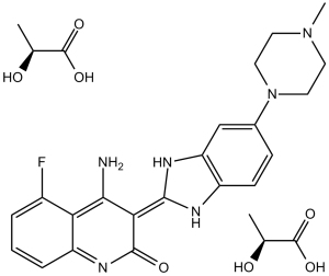
| Formula | C21H21FN6O.2C3H6O3 |
| CAS No. | 852433-84-2 |
| Storage | -20℃ for 3 years in powder form |
| -80℃ for 2 years in solvent | |
| Solubility (In vitro) | DMSO: 90 mg/mL (157.2 mM) |
| Water: 70 mg/mL (122.3 mM) | |
| Ethanol:<1 mg/mL | |
| Solubility (In vivo) | Saline: 30 mg/kg |
| Synonyms | TKI-258, CHIR-258; TKI258; TKI 258; CHIR258; CHIR 258; TKI258 Dilactic acid |
| In Vitro | In vitro activity: Dovitinib potently inhibits the FGF-stimulated growth of WT and F384L-FGFR3-expressing B9 cells with IC50 of 25 nM. In addition, Dovitinib inhibits proliferation of B9 cells expressing each of the various activated mutants of FGFR3. Interestingly, there are minimal observed differences in the sensitivity of the different FGFR3 mutations to Dovitinib, with the IC50 ranging from 70 to 90 nM for each of the various mutations. IL-6-dependent B9 cells containing vector only (B9-MINV cells are resistant to the inhibitory activity of Dovitinib at concentrations up to 1 μM. Dovitinib inhibits cell proliferation of KMS11 (FGFR3-Y373C), OPM2 (FGFR3-K650E), and KMS18 (FGFR3-G384D) cells with IC50 of 90 nM (KMS11 and OPM2) and 550 nM, respectively. Dovitinib inhibits FGF-mediated ERK1/2 phosphorylation and induces cytotoxicity in FGFR3-expressing primary MM cells. BMSCs does confer a modest degree of resistance with 44.6% growth inhibition for cells treated with 500 nM Dovitinib and cultured on stroma compared with 71.6% growth inhibition for cells grown without BMSCs. Dovitinib inhibits proliferation of M-NFS-60, an M-CSF growth-driven mouse myeloblastic cell line with a median effective concentration (EC50) of 220 nM. Treatment of SK-HEP1 cells with Dovitinib results in a dose-dependent reduction in cell number and G2/M phase arrest with reduction in the G0/G1 and S phases, inhibition of anchorage-independent growth and blockage of bFGF-induced cell motility. The IC50 for Dovitinib in SK-HEP1 cells is approximately 1.7 μM. Dovitinib also significantly reduces the basal phosphorylation levels of FGFR-1, FGFR substrate 2α (FRS2-α) and ERK1/2 but not Akt in both SK-HEP1 and 21-0208 cells. In 21-0208 HCC cells, Dovitinib significantly inhibits bFGF-induced phosphorylation of FGFR-1, FRS2-α, ERK1/2 but not Akt. Kinase Assay: The inhibitory concentration of 50% (IC50) values for the inhibition of RTKs by Dovitinib are determined in a time-resolved fluorescence (TRF) or radioactive format, measuring the inhibition by Dovitinib of phosphate transfer to a substrate by the respective enzyme. The kinase domains of FGFR3, FGFR1, PDGFRβ, and VEGFR1-3 are assayed in 50 mM HEPES (N-2-hydroxyethylpiperazine-N′-2-ethanesulfonic acid), pH 7.0, 2 mM MgCl2, 10 mM MnCl2 1 mM NaF, 1 mM dithiothreitol (DTT), 1 mg/mL bovine serum albumin (BSA), 0.25 μM biotinylated peptide substrate (GGGGQDGKDYIVLPI), and 1 to 30 μM adenosine triphosphate (ATP) depending on the Km for the respective enzyme. ATP concentrations are at or just below Km. For c-KIT and FLT3 reactions the pH is raised to 7.5 with 0.2 to 8 μM ATP in the presence of 0.25 to 1 μM biotinylated peptide substrate (GGLFDDPSYVNVQNL). Reactions are incubated at room temperature for 1 to 4 hours and the phosphorylated peptide captured on streptavidin-coated microtiter plates containing stop reaction buffer (25 mM EDTA [ethylenediaminetetraacetic acid], 50 mM HEPES, pH 7.5). Phosphorylated peptide is measured with the DELFIA TRF system using a Europium-labeled antiphosphotyrosine antibody PT66. The concentration of Dovitinib for IC50 is calculated using nonlinear regression with XL-Fit data analysis software version 4.1 (IDBS). Inhibition of colony-stimulating factor-1 receptor (CSF-1R), PDGFRα, insulin receptor (InsR), and insulin-like growth factor receptor 1 (IGFR1) kinase activity is determined at ATP concentrations close the Km for ATP. Cell Assay: Cell viability is assessed by 3-(4,5-dimethylthiazol)-2,5-diphenyl tetrazolium (MTT) dye absorbance. Cells are seeded in 96-well plates at a density of 5 × 10³ (B9 cells) or 2 × 10⁴ (MM cell lines) cells per well. Cells are incubated with 30 ng/mL aFGF and 100 μg/mL heparin or 1% IL-6 where indicated and increasing concentrations of Dovitinib. For each concentration of Dovitinib, 10 μL aliquots of drug or DMSO diluted in culture medium is added. For drug combination studies, cells are incubated with 0.5 μM dexamethasone, 100 nM Dovitinib, or both simultaneously where indicated. To evaluate the effect of Dovitinib on growth of MM cells adherent to BMSCs, 104 KMS11 cells are cultured on BMSC-coated 96-well plates in the presence or absence of Dovitinib. Plates are incubated for 48 to 96 hours. For assessment of macrophage colony-stimulating factor (M-CSF)-mediated growth, 5 × 10³ M-NFS-60 cells/well are incubated with serial dilutions of Dovitinib with 10 ng/mL M-CSF and without granulocyte-macrophage colony-stimulating factor (GM-CSF). After 72 hours cell viability is determined using Cell Titer-Glo Assay. Each experimental condition is performed in triplicate. |
|---|---|
| In Vivo | Dovitinib induces both cytostatic and cytotoxic responses in vivo resulting in regression of FGFR3-expressing tumors. Dovitinib shows a dose- and exposure-dependent inhibition of target receptor tyrosine kinases (RTKs) expressed in tumor xenografts. Dovitinib potently inhibits tumor growth of six HCC lines. Inhibition of angiogenesis correlated with inactivation of FGFR/PDGFRβ/VEGFR2 signaling pathways. In an orthotopic model, Dovitinib potently inhibits primary tumor growth and lung metastasis and significantly prolonged mouse survival. Administration of Dovitinib results in significant tumor growth inhibition and tumor regressions, including large, established tumors (500-1,000 mm3). |
| Animal model | Female BNX mice bearing KMS11 cells |
| Formulation & Dosage | Dissolved in 5 mM citrate buffer; 10, 30, or 60 mg/kg; p.o. |
| References | Blood. 2005 Apr 1;105(7):2941-8; J Hepatol. 2012 Mar;56(3):595-601; Clin Cancer Res. 2005 May 15;11(10):3633-41. |
 m.cnreagent.com