| CAS NO: | 215543-92-3 |
| 规格: | ≥98% |
| 包装 | 价格(元) |
| 5mg | 电议 |
| 25mg | 电议 |
| 50mg | 电议 |
| 100mg | 电议 |
| 250mg | 电议 |
| 500mg | 电议 |
| 1g | 电议 |
| Molecular Weight (MW) | 296.32 |
|---|---|
| Formula | C17H16N2O3 |
| CAS No. | 215543-92-3 |
| Storage | -20℃ for 3 years in powder form |
| -80℃ for 2 years in solvent | |
| Solubility (In vitro) | DMSO: 59 mg/mL (199.1 mM) |
| Water: <1 mg/mL | |
| Ethanol:<1 mg/mL | |
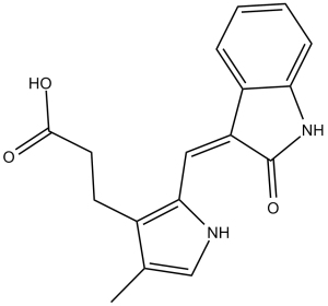
| Other info | Chemical Name: (Z)-3-(4-methyl-2-((2-oxoindolin-3-ylidene)methyl)-1H-pyrrol-3-yl)propanoic acid. InChi Key: JNDVEAXZWJIOKB-JYRVWZFOSA-N InChi Code: InChI=1S/C17H16N2O3/c1-10-9-18-15(11(10)6-7-16(20)21)8-13-12-4-2-3-5-14(12)19-17(13)22/h2-5,8-9,18H,6-7H2,1H3,(H,19,22)(H,20,21)/b13-8- SMILES Code: O=C(O)CCC1=C(/C=C2C(NC3=C\2C=CC=C3)=O)NC=C1C |
| Synonyms | SU-5402; SU 5402; SU5402. |
| In Vitro | In vitro activity: SU5402 inhibits VEGF-, FGF-, PDGF- dependent cell proliferation with IC50 of 0.05 μM, 2.80μM, 28.4 μM, respectively. In HUVECs, SU5416 selectively inhibits VEGF-driven mitogenesis in a dose-dependent manner with IC50 of 0.04 μM. In nasopharyngeal epithelial cells, SU5402 attenuates LMP1-mediated aerobic glycolysis, cellular transformation, cell migration, and invasion. In mouse C3H10T1/2 cells, SU 5402 diminishes the effect of FGF23 on cell differentiation Kinase Assay: The catalytic portion of FGF-R1 and Flk-1/KDR are expressed as GST fusion proteins following infection of Spodoptera frugiperda (sf9) cells with engineered baculoviruses. GST-FGFR1 and GST-Flk1 are purified to homogeneity from infected sf9 cell lysates by glutathione sepharose chromatography. The assays are performed in 96-well microtiter plates that had been coated overnight with 2.0 μg of a polyGlu-Tyr peptide (4:1) in 0.1 mL of PBS per well. The purified kinases are diluted in kinase assay buffer (100 mM Hepes pH 7.5, 100 mM NaCl, and 0.1 mM sodium orthovanadate) and added to all test wells at 5 ng of GST fusion protein per 0.05 mL volume buffer. Test compounds are diluted in 4% DMSO and added to test wells (0.025 mL/well). The kinase reaction is initiated by the addition of 0.025 mL of 40 μM ATP/40 mM MnCl2, and plates are shaken for 10 min before stopping the reactions with the addition of 0.025 mL of 0.5 M EDTA. The final ATP concentration was 10 μM, which is twice the experimentally determined Km value for ATP. Negative control wells receive MnCl2 alone without ATP. The plates are washed three times with 10 mM Tris pH 7.4, 150 mM NaCl, and 0.05% Tween-20 (TBST). Rabbit polyclonal anti-phosphotyrosine antiserum is added to the wells at a 1:10000 dilution in TBST for 1 h. The plates are then washed three times with TBST. Goat anti-rabbit antiserum conjugated with horseradish peroxidase was then added to all wells for 1 h. The plates are washed three times with TBST, and the peroxidase reaction is detected with the addition of 2,2‘-azinobis(3-ethylbenzthiazoline-6-sulfonic acid) (ABTS). The color readout of the assay is allowed to develop for 20–30 min and read on a Dynatech MR5000 ELISA plate reader using a 410 nM test filter. Cell Assay: Tumor cell lines (SF767T, SF763, EPH4-VEGF, C6, A375, A431, LNCAP, Calu-6, 3T3Her2 and 488G2M2 cells) used in the in vitro growth are cultured in media at 37°C in 5–10% CO2. SU5416 is serially diluted in media containing DMSO (<0.5%) and added to cultures of tumor cells 1 day after the initiation of culture. Cell growth is measured after 96 h using the sulforhodamine B method. IC50s are calculated by curve fitting using four-parameter analysis. |
|---|---|
| In Vivo | In mice, SU5416 (25 mg/kg, i.p.) inhibits subcutaneous growth of a panel of tumor cell lines by inhibiting the angiogenic process associated with tumor growth. |
| Animal model | Mice bearing SF767T, SF763, EPH4-VEGF, C6, A375, A431, LNCAP, Calu-6, 3T3Her2 or 488G2M2 tumors |
| Formulation & Dosage | Dissolved in DMSO; 25mg/kg; i.p. injection |
| References | J Med Chem. 1999 Dec 16;42(25):5120-30; Cancer Res. 1999 Jan 1;59(1):99-106. |
NIH 3T3 Flk-1 cells (A) or NIH 3T3 platelet-derived growth factor β cells (B) grown to confluency were preincubated with SU5416 at concentrations ranging from 0.05 to 50 μm for 1 h at 37°C. Cancer Res. 1999 Jan 1;59(1):99-106. |
A375 cells (3 × 10⁶) were implanted subcutaneous into the hindflank region of female BALB/c nu/nu mice 8–12 weeks of age. Cancer Res. 1999 Jan 1;59(1):99-106. |
Rat C6 glioma cells were surgically implanted (0.5 × 10⁶ cells/animal) under the serosa of the colon in BALB/c nu/nu mice. Beginning 1 day after implantation, animals were treated once daily with a 50 μl i.p. bolus injection of either SU5416 at 25 mg/kg/day in DMSO or DMSO alone for 16 days. On day 16 after implantation, animals were euthanized, and their local tumors in the colon were first quantitated by measurement using venier calipers and then harvested. Cancer Res. 1999 Jan 1;59(1):99-106. |
 m.cnreagent.com

