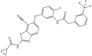
| CAS NO: | 1228591-30-7 |
| 规格: | ≥98% |
| 包装 | 价格(元) |
| 10mg | 电议 |
| 25mg | 电议 |
| 50mg | 电议 |
| 100mg | 电议 |
| 250mg | 电议 |
| 500mg | 电议 |
| Molecular Weight (MW) | 554.52 |
|---|---|
| Formula | C27H18F4N4O3S |
| CAS No. | 1228591-30-7 |
| Storage | -20℃ for 3 years in powder form |
| -80℃ for 2 years in solvent | |
| Solubility (In vitro) | DMSO: 100 mg/mL (180.3 mM) |
| Water: <1 mg/mL | |
| Ethanol: 2 mg/mL (3.6 mM) | |
| Solubility (In vivo) | 2% DMSO+98% PEG 300: 5 mg/mL |
| Synonyms | TAK632, TAK 632, TAK-632; N-(7-cyano-6-(4-fluoro-3-(2-(3-(trifluoromethyl)phenyl)acetamido)phenoxy)benzo[d]thiazol-2-yl)cyclopropanecarboxamide |
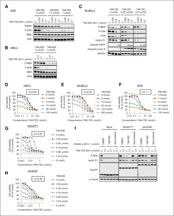
| In Vitro | In vitro activity: TAK-632 inhibits phosphorylation of MEK and ERK in melanoma A375 cells (BRAFV600E) with IC50 of 12 nM and 16 nM, respectively. In human melanoma HMVII cells (NRASQ61K/BRAFG469V), TAK-632 also shows strong inhibition of pMEK and pERK with IC50 of 49 nM and 50 nM, respectively. Moreover, TAK-632 also exhibits antiproliferative activity in both A375 and HMVII cells with GI50 of 66 nM and 200 nM, respectively. TAK-632 induces RAF dimerization but inhibits the kinase activity of the RAF dimer because of its slow dissociation from RAF. The combination of TAK-632 and TAK-733 exhibits synergistic antiproliferative effects in BRAF- and NRAS-mutated melanoma cells. Kinase Assay: Assays for serine/threonine kinases using radio labeled [γ-33P] ATP are performed in 96 well plates. BRAF and c-RAF are expressed as N-terminal FLAG-tagged protein using a baculovirus expression system. The reaction conditions are optimized for each kinase: BRAF (25 ng/well of enzyme, 1 μg/well of GST-MEK1(K96R), 0.1 μCi/well of [γ-32P] ATP, room temperature, 20 min reaction); c-RAF (25 ng/well of enzyme, 1 μg/well of GST-MEK1 (K96R), 0.1 μCi/well of [γ-32P] ATP, room temperature, 20 min reaction). Enzyme reactions are performed in 25 mM HEPES, pH 7.5, 10 mM magnesium acetate, 1 mM dithiothreitol and 0.5 μM ATP containing optimized concentration of enzyme, substrate and radiolabeled ATP as described above in a total volume of 50 μL. Prior to the kinase reaction, compound and enzyme are incubated for 5 min at reaction temperature as described above. The kinase reactions are initiated by adding ATP. After the reaction period as described above, the reactions are terminated by the addition of 10% (final concentration) trichloroacetic acid. The [γ-33P] or [γ-32P]-phosphorylated proteins are filtered in GFC filter plates with a Cell Harvester and then the plates are washed out with 3% phosphoric acid. The plates are dried, followed by the addition of 40 μL of MicroScint0. The radioactivity is counted by a TopCount scintillation counter. Cell Assay: The cells (A375 and HMVII cells) are proliferated in appropriate medium (vender recommended) supplemented with 10% heat-inactivated fetal bovine serum (FBS) and antibiotics, in tissue culture dishes placed in a humidified incubator maintained at 37°C in an atmosphere of 5% CO2 and 95% air. The anti-proliferative activity of compound is determined by treating cell lines with the compound for 3 days, followed by measurement of viable cell number in the Cell Titer-Glo assay. The cells are seeded in a 96-multiwell plate at 1500 to 4000 cells per well in medium containing FBS and cells allowed to sit down overnight. After 18–20 h, compounds at various concentrations by serial dilution are added to the cells and were cultured for 3 days in chamber. After the treatment culture, cellular proliferation is determined by a Cell Titer-Glo Luminescent Cell Viability Assay. In brief, 100 bL/well of Cell Titer-Glo Substrate solution is added to each well and the cells were cultured for an additional 10 minutes (approximately). The chemi-luminescence value is measured using a Luminescence Counter 1420 ARVO MX Light. Concentration response curves are generated by calculating the decrease in chemi-luminescence values in compound-treated samples relative to the vehicle (DMSO) treated controls. |
|---|---|
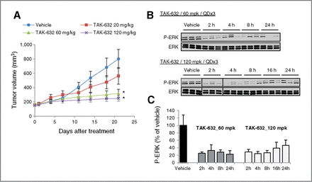
| In Vivo | TAK-632 shows superior oral bioavailability in both rats and dogs. TAK-632 (3.9–24.1 mg/kg, p.o.) exhibits dose-dependent antitumor efficacy without severe body weight reduction in a melanoma A375 (BRAFV600E) xenograft model and a human melanoma HMVII (NRASQ61K/BRAFG469V) xenograft in rats. In NRAS-mutant melanoma SK-MEL-2 xenograft model, TAK-632 (60 or 120 mg/kg, p.o.) also exhibits potent antitumor efficacy without severe toxicity. |
| Animal model | Human melanoma A375 (BRAFV600E) xenograft model and human melanoma HMVII (NRASQ61K/BRAFG469V) xenograft model in rats. |
| Formulation & Dosage | Dissolved in hydroxypropyl methylcellulose phthalate and distilled water; 24.1mg/kg; Oral gavage |
| References | J Med Chem. 2013 Aug 22;56(16):6478-94; Cancer Res. 2013 Dec 1;73(23):7043-55. |
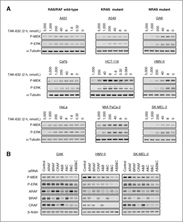
Effect of TAK-632 on MAPK pathway. Cancer Res. 2013 Dec 1;73(23):7043-55. |
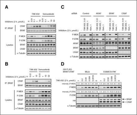
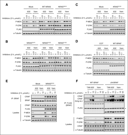
Formation of RAF dimer mediates RAF paradoxical activation by TAK-632. A and B, SK-MEL-2 and A549 cells were treated with TAK-632 and vemurafenib at the indicated concentrations for 2 hours, respectively. Cancer Res. 2013 Dec 1;73(23):7043-55. |
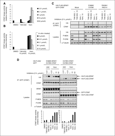
TAK-632 suppresses the kinase activity of RAF dimer. A, SK-MEL-2 cells were treated with TAK-632 or vemurafenib (vem) at indicated concentrations for 2 hours. Cancer Res. 2013 Dec 1;73(23):7043-55. |
Effect of TAK-632 on xenograft proliferation. A, mice bearing SK-MEL-2 xenografts were treated once daily for 21 consecutive days with vehicle or TAK-632 SD at the indicated concentrations (10 mice per each treatment group). Cancer Res. 2013 Dec 1;73(23):7043-55. |
TAK-632 suppresses MAPK pathway in vemurafenib-resistant melanoma cells. Cancer Res. 2013 Dec 1;73(23):7043-55. |
TAK-632 shows synergy with a MEK inhibitor. Cancer Res. 2013 Dec 1;73(23):7043-55. |
 m.cnreagent.com