| CAS NO: | 152121-30-7 |
| 规格: | ≥98% |
| 包装 | 价格(元) |
| 5mg | 电议 |
| 10mg | 电议 |
| 25mg | 电议 |
| 50mg | 电议 |
| 100mg | 电议 |
| 250mg | 电议 |
| 500mg | 电议 |
| 1g | 电议 |
| Molecular Weight (MW) | 331.34 |
|---|---|
| Formula | C20H14N3OF |
| CAS No. | 152121-30-7 |
| Storage | -20℃ for 3 years in powder form |
| -80℃ for 2 years in solvent | |
| Solubility (In vitro) | DMSO: 66 mg/mL (199.2 mM) |
| Water: <1 mg/mL | |
| Ethanol: 12 mg/mL (36.2 mM) | |
| Solubility (In vivo) | 1% DMSO+30% polyethylene glycol+1% Tween 80: 30 mg/mL |
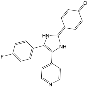
| Synonyms | FHPI; SB-202190; SB202190; SB202190 Chemical Name: 4-(4-(4-fluorophenyl)-5-(pyridin-4-yl)-1H-imidazol-2-yl)phenol SMILES Code: OC1=CC=C(C2=NC(C3=CC=C(F)C=C3)=C(C4=CC=NC=C4)N2)C=C1 |
| In Vitro | In vitro activity: SB 202190 significantly inhibits both basal and anti-Fas antibody-induced MAPKAPK 2 activity in a dose-dependent manner. SB202190 by itself is sufficient to induce cell death in Jurkat and HeLa cells through activation of CPP32-like caspases, which can be blocked by expression of bcl-2. SB202190-induced apoptosis is attenuated by p38β but augmented by p38α. SB 202190 strongly inhibits UVB induced COX-2 protein expression in HaCaT cells, and markedly inhibits UVB induced cox-2 mRNA. SB 202190 treatment inhibits the expression of albumin-induced proinflammatory (monocyte chemoattractant protein-1) and transforming growth factor (TGF)-beta1-induced profibrotic (procollagen-Ialpha1) genes over 50% in renal tubular cells (normal rat kidney-52E). Kinase Assay: The p38α and p38β are assayed in 25 mM Tris-HCl, pH 7.5, containing 0.1 mM EGTA, with myelin basic protein (0.33 mg/mL) as substrate. Assays are performed either manually for 10 minutes at 30 °C in 50 μL incubations using [γ-33P]ATP, or with a Biomek 2000 Laboratory Automation Workstation in a 96-well format for 40 minutes at ambient temperature in 25 μL incubations using [γ-33P]ATP. The concentrations of ATP and magnesium acetate are 0.1 mM and 10 mM respectively. All assays are initiated with MgATP. Manual assays are terminated by spotting aliquots of incubation on to phosphocellulose paper, followed by immersion in 50 mM phosphoric acid. Robotic assays are terminated by the addition of 5 μL of 0.5 M phosphoric acid before spotting aliquots on to P30 filter mats. All papers are then washed four times in 50 mM phosphoric acid to remove ATP, once in acetone (manual incubations) or methanol (robotic incubations), and then dried and counted for radioactivity. Cell Assay: Cells are serum-starved and then treated with different concentration of SB 202190 for 24 hours. Cell viability is assayed by either trypan blue exclusion or propidium iodide exclusion followed by flow cytometry analysis. The apoptotic nuclei are visualized by H33258 staining. |
|---|---|
| In Vivo | Inhibiting p38 by administration of SB 202190 inhibits PV IgG-induced blister formation in the passive transfer mouse model. In the endotoxin model of sepsis, SB 202190 treatment produces a statistically significant survival benefit compared with control. |
| Animal model | C57BL/6J mice injected with IgG or PV IgG |
| Formulation & Dosage | Dissolved in DMSO and diluted in saline; 12.5 μg; Administered via i.d. |
| References | Biochem J. 2000 Oct 1;351(Pt 1):95-105; Proc Natl Acad Sci U S A. 2006 Aug 22;103(34):12855-60; J Surg Res. 2009 Mar;152(1):46-53. |
Inhibiting p38MAPK prevents clinical blistering in PV passive transfer mice. Neonatal C57BL/6J mice were injected i.d. with either PV IgG (1.5 mg of IgG/g body weight) (Left) or PV IgG (1.5 mg of IgG/g body weight) plus SB202190 (Right). After 18 h, the skin of neonatal mice from the test and control groups was examined clinically. PV IgG-treated mice have a positive Nikolsky’s sign (white arrows), demonstrating loss of epithelial cell–cell adhesion. In contrast, mice treated with the SB202190 and PV IgG have a negative Nikoslky’s sign, indicating that epithelial adhesion remains intact. Proc Natl Acad Sci U S A. 2006 Aug 22; 103(34): 12855–12860. |
Inhibition of PV IgG-mediated p38MAPK and HSP27 phosphorylation in skin of PV IgG plus SB202190-treated mice. Neonatal C57BL/6 WT mice were injected i.d. with control IgG (CON; 1 mg of IgG/g body weight), PV IgG (1 mg of IgG/g body weight), or SB202190 and then PV IgG (PV IgG plus SB202190). Skin biopsies were obtained after 18 h of treatment and extracted in IEF lysis buffer. Proc Natl Acad Sci U S A. 2006 Aug 22; 103(34): 12855–12860. |
 m.cnreagent.com
