| CAS NO: | 942918-07-2 |
| 规格: | ≥98% |
| 包装 | 价格(元) |
| 5mg | 电议 |
| 25mg | 电议 |
| 50mg | 电议 |
| 100mg | 电议 |
| 250mg | 电议 |
| 500mg | 电议 |
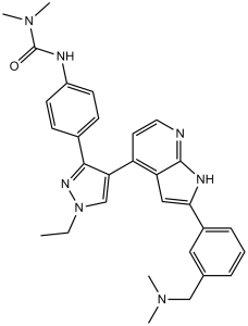
| Molecular Weight (MW) | 507.63 |
|---|---|
| Formula | C30H33N7O |
| CAS No. | 942918-07-2 |
| Storage | -20℃ for 3 years in powder form |
| -80℃ for 2 years in solvent | |
| Solubility (In vitro) | DMSO: 102 mg/mL (200.9 mM) |
| Water: <1 mg/mL | |
| Ethanol: N/a | |
| Solubility (In vivo) | 2% Cremophor EL, 2% N,N-dimethylacetamide, pH 5.0: ~30 mg/mL |
| Synonyms | GSK-1070916A; GSK 1070916A; GSK1070916A; GSK-1070916; GSK 1070916; GSK1070916; NMI 900; NMI900; NMI-900. |
| In Vitro | In vitro activity: GSK1070916 selectively inhibits Aurora B and Aurora C with Ki of 0.38 nM and 1.5 nM over Aurora A with Ki of 490 nM. Inhibition of Aurora B and Aurora C is time-dependent, with an enzyme-inhibitor dissociation half-life of>480 min and 270 min respectively. In addition, GSK1070916 is also a competitive inhibitor with respect to ATP. Human tumor cells treated with GSK1070916 shows dose-dependent inhibition of phosphorylation on serine 10 of Histone H3, a substrate specific for Aurora B. Moreover, GSK1070916 inhibits the proliferation of tumor cells with EC50 values of<10 nM in over 100 cell lines spanning a broad range of tumor types, with a median EC50 of 8 nM. Although GSK1070916 has potent activity against proliferating cells, a dramatic shift in potency is observed in primary, nondividing, normal human vein endothelial cells. Furthermore, GSK1070916-treated cells do not arrest in mitosis but instead fails to divide and become polyploid, ultimately leading to apoptosis. In another study, it is also reported high chromosome number associated with resistance to the inhibition of Aurora B and C suggests cells with a mechanism to bypass the high ploidy checkpoint are resistant to GSK1070916. Kinase Assay: The ability of GSK1070916 to inhibit the Aurora enzymes is measured using in vivo kinase assays. The assays measure the ability of Aurora A, Aurora B and Aurora C to phosphorylate a synthetic peptide substrate. Biotin-Ahx-RARRRLSFFFFAKKK-NH2 is used for the Aurora A–TPX2 LEADseekerTM assay and 5FAM-PKAtide is used for the IMAPTM assay for all three Aurora kinases. To take into account time-dependent inhibition of Aurora enzymes, Aurora A–TPX2, Aurora B–INCENP and Aurora C–INCENP are incubated with GSK1070916 at various concentrations for 30 min before the reactions are initiated with the addition of substrates. For the Aurora A LEADseekerTM assay, final assay conditions are 0.5 nM Aurora A–TPX2, 1 μM peptide substrate, 6 mM MgCl2, 1.5 μM ATP, 0.003 μCi/μL [γ-33P] ATP in 50 mM Hepes, pH 7.2, 0.15 mg/mL BSA, 0.01% Tween-20, 5 mM DTT and 25 mM KCl. The reactions are incubated at room temperature (25 °C) for 120 min and terminated by the addition of LEADseekerTM beads in PBS containing EDTA (final concentration 2 mg/mL beads and 25 mM EDTA). The plates are then sealed, and the beads are allowed to settle overnight. Product formation is quantified using a Viewlux Imager. For the IMAPTM assays, Aurora A–TPX2 (final concentration 1 nM), Aurora B–INCENP (final concentration 2 nM) or Aurora C–INCENP (final concentration 2.5 nM) is added to the compound-containing plates in 5 μL of buffer (25 mM Hepes, pH 7.2, for Aurora A, 25 mM Hepes, pH 7.5, for Aurora B and 20 mM Hepes, pH 7.2, for Aurora C) containing 0.15 mg/mL BSA, 0.01% Tween 20 and 25 mM NaCl. This mixture is incubated at room temperature for 30 min. To start the reaction, 5 μL of a substrate solution is added containing the same Hepes buffer as used for the pre-incubation, 25 mM NaCl, MgCl2 (2, 4 and 4 mM for Aurora A, B and C respectively), DTT (4, 4 and 2 mM for Aurora A, B and C respectively), ATP (4, 4 and 10 μM for Aurora A, B and C respectively), 200 nM 5FAM-PKAtide, 0.01% Tween 20 and 0.15 mg/mL BSA. The reactions are incubated at room temperature for 120 min for Aurora A and B and 60 min for Aurora C. These reactions are then terminated by the addition of 10 μL of 1:500 (1:600 for Aurora C) Progressive Binding Reagent in 95% Progressive Binding Buffer A and 5% Progressive Binding Buffer B. Plates are incubated at room temperature for approx. 90–120 min (time allowed for equilibrium to be reached). Plates are read in a Molecular Devices Analyst plate reader in fluorescence polarization mode. Cell Assay: The ability of GSK1070916 to inhibit the Aurora enzymes is measured using in vivo kinase assays. The assays measure the ability of Aurora A, Aurora B and Aurora C to phosphorylate a synthetic peptide substrate. Biotin-Ahx-RARRRLSFFFFAKKK-NH2 is used for the Aurora A–TPX2 LEADseekerTM assay and 5FAM-PKAtide is used for the IMAPTM assay for all three Aurora kinases. To take into account time-dependent inhibition of Aurora enzymes, Aurora A–TPX2, Aurora B–INCENP and Aurora C–INCENP are incubated with GSK1070916 at various concentrations for 30 min before the reactions are initiated with the addition of substrates. For the Aurora A LEADseekerTM assay, final assay conditions are 0.5 nM Aurora A–TPX2, 1 μM peptide substrate, 6 mM MgCl2, 1.5 μM ATP, 0.003 μCi/μL [γ-33P] ATP in 50 mM Hepes, pH 7.2, 0.15 mg/mL BSA, 0.01% Tween-20, 5 mM DTT and 25 mM KCl. The reactions are incubated at room temperature (25 °C) for 120 min and terminated by the addition of LEADseekerTM beads in PBS containing EDTA (final concentration 2 mg/mL beads and 25 mM EDTA). The plates are then sealed, and the beads are allowed to settle overnight. Product formation is quantified using a Viewlux Imager. For the IMAPTM assays, Aurora A–TPX2 (final concentration 1 nM), Aurora B–INCENP (final concentration 2 nM) or Aurora C–INCENP (final concentration 2.5 nM) is added to the compound-containing plates in 5 μL of buffer (25 mM Hepes, pH 7.2, for Aurora A, 25 mM Hepes, pH 7.5, for Aurora B and 20 mM Hepes, pH 7.2, for Aurora C) containing 0.15 mg/mL BSA, 0.01% Tween 20 and 25 mM NaCl. This mixture is incubated at room temperature for 30 min. To start the reaction, 5 μL of a substrate solution is added containing the same Hepes buffer as used for the pre-incubation, 25 mM NaCl, MgCl2 (2, 4 and 4 mM for Aurora A, B and C respectively), DTT (4, 4 and 2 mM for Aurora A, B and C respectively), ATP (4, 4 and 10 μM for Aurora A, B and C respectively), 200 nM 5FAM-PKAtide, 0.01% Tween 20 and 0.15 mg/mL BSA. The reactions are incubated at room temperature for 120 min for Aurora A and B and 60 min for Aurora C. These reactions are then terminated by the addition of 10 μL of 1:500 (1:600 for Aurora C) Progressive Binding Reagent in 95% Progressive Binding Buffer A and 5% Progressive Binding Buffer B. Plates are incubated at room temperature for approx. 90–120 min (time allowed for equilibrium to be reached). Plates are read in a Molecular Devices Analyst plate reader in fluorescence polarization mode. |
|---|---|
| In Vivo | GSK1070916 (25, 50, or 100 mg/kg) shows dose-dependent inhibition of phosphorylation of an Aurora B–specific substrate in mice and consistent with its broad cellular activity, has antitumor effects in 10 human tumor xenograft models including breast, colon, lung, and two leukemia models. |
| Animal model | Mice tumor xenograft models (A549, SW620, HCT116, H460, MCF-7, HL60, K562, Colo205) |
| Formulation & Dosage | Formulated in 2% Cremophor EL, 2% N,N-dimethylacetamide, and 96% acidified water (pH 5.0); 25, 50, or 100 mg/kg; i.p. injection. |
| References | Biochem J. 2009 May 13;420(2):259-65; Mol Cancer Ther. 2009 Jul;8(7):1808-17. |
 m.cnreagent.com