| CAS NO: | 443797-96-4 |
| 规格: | ≥98% |
| 包装 | 价格(元) |
| 2mg | 电议 |
| 5mg | 电议 |
| 10mg | 电议 |
| 25mg | 电议 |
| 50mg | 电议 |
| 100mg | 电议 |
| 250mg | 电议 |
| 500mg | 电议 |
| Molecular Weight (MW) | 394.36 |
|---|---|
| Formula | C15H12F2N6O3S |
| CAS No. | 443797-96-4 |
| Storage | -20℃ for 3 years in powder form |
| -80℃ for 2 years in solvent | |
| Solubility (In vitro) | DMSO: 79 mg/mL (200.3 mM) |
| Water: <1 mg/mL | |
| Ethanol:<1 mg/mL | |
| Solubility (In vivo) | 0.5% methylcellulose+0.2% Tween 80: 14 mg/mL |
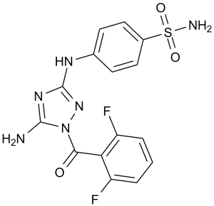
| Other info | Synonym: JNJ 7706621; JNJ-7706621; JNJ7706621 Chemical Name: 4-((5-amino-1-(2,6-difluorobenzoyl)-1H-1,2,4-triazol-3-yl)amino)benzenesulfonamide InChi Key: KDKUVYLMPJIGKA-UHFFFAOYSA-N InChi Code: InChI=1S/C15H12F2N6O3S/c16-10-2-1-3-11(17)12(10)13(24)23-14(18)21-15(22-23)20-8-4-6-9(7-5-8)27(19,25)26/h1-7H,(H2,19,25,26)(H3,18,20,21,22) SMILES Code: O=S(C1=CC=C(NC2=NN(C(C3=C(F)C=CC=C3F)=O)C(N)=N2)C=C1)(N)=O |
| In Vitro | In vitro activity: JNJ-7706621 also shows some inhibition to VEGF-R2, FGF-R2, and GSK3β, with IC50 of 154-254 nM. JNJ-7706621 shows inhibitory effect on a panel of human cancer cell types, including HeLa, HCT-116, SK-OV-3, PC3, DU145, A375, MDA-MB-231, MES-SA, and MES-SA/Dx5, with IC50 of 112-514 nM, independent of p53, retinoblastoma, or P-glycoprotein status. JNJ-7706621 is several-fold less potent at inhibiting growth of normal cell types, including MRC-5, HASMC, HUVEC, and HMVEC, with IC50 of 3.67-5.42 μM. In HeLa or U937 cells, JNJ-7706621 (0.5-3 μM) delays exit from G1, arrests cells in G2-M, induces endoreduplication, activates apoptosis, and reduces colony formation. In a HeLa cell line, incremental treatment with increasing concentrations of JNJ-7706621 leads to a 16-fold resistance, which may be mediated by ABCG2. Kinase Assay: For CDK1 kinase activity, a method is developed using the CDK1/cyclin B complex purified from baculovirus to phosphorylate a biotinylated peptide substrate containing the consensus phosphorylation site for histone H1, which is phosphorylated in vivo by CDK1. Inhibition of CDK1 activity is measured by observing a reduced amount of 33P-γ-ATP incorporation into the immobilized substrate in streptavidin-coated 96-well scintillating microplates. CDK1 enzyme is diluted in 50 mM Tris-HCl (pH 8), 10 mM MgCl2, 0.1 mM Na3VO 4, 1 mM DTT, 1% DMSO, 0.25 μM peptide, 0.1 μCi per well 33P-γ-ATP, and 5 μM ATP in the presence or absence of various concentrations of JNJ-7706621 and incubated at 30 °C for 1 hour. The reaction is terminated by washing with PBS containing 100 mM EDTA and plates are counted in a scintillation counter. Linear regression analysis of the percent inhibition by JNJ-7706621 is used to determine IC50. The Aurora kinase assays are done with 10 μM ATP and a peptide containing a dual repeat of the kemptide phosphorylation motif. Cell Assay: The ability of JNJ-7706621 to inhibit the proliferation of cell growth is determined by measuring incorporation of 14C-labelled thymidine into newly synthesized DNA within the cells. Cells are trypsinized and counted and 3-8 × 10³ cells are added to each well of a 96-well CytoStar tissue culture treated scintillating microplate in complete medium in a volume of 100 μL. Cells are incubated for 24 hours in complete medium at 37 °C in an atmosphere containing 5% CO2. Next, 1 μL of JNJ-7706621 is added to the wells of the plate. Cells are incubated for 24 more hours. Methyl 14C-thymidine 56 mCi/mmol is diluted in complete medium and 0.2 μCi/well is added to each well of the CytoStar plate in a volume of 20 μL. The plate is incubated for 24 hours at 37 °C in JNJ-7706621 plus 14C-thymidine. The contents of the plate are discarded and the plate is washed twice with 200 μL PBS. 200 μL of PBS is added to each well. The top of the plate is sealed with a transparent plate sealer and a white plate backing sealer is applied to the bottom of the plate. The degree of methyl 14C-thymidine incorporation is quantified on a Packard Top Count. |
|---|---|
| In Vivo | In mouse xenograft model of A375 melanoma human tumor, JNJ-7706621 (100 or 125 mg/kg) causes tumor regression. |
| Animal model | Mouse xenograft model of A375 cells |
| Formulation & Dosage | Dissolved in 0.5% methylcellulose containing 0.1% polysorbate 80 in sterile water; 100, 125 mg/kg; oral administration or i.p. injection. |
| References | Cancer Res. 2005 Oct 1;65(19):9038-46; Mol Cancer Ther. 2006 Oct;5(10):2459-67; J Med Chem. 2005 Jun 30;48(13):4208-11. |
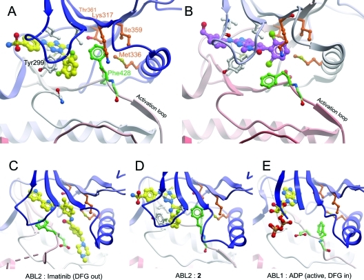
ABL2 bound to a type I inhibitor 2. (A) ABL2:2, showing the compound bound to the ATP binding site, and the ordered activation loop. Compound 2 is shown in yellow. J Med Chem. 2011 Apr 14;54(7):2359-67. |
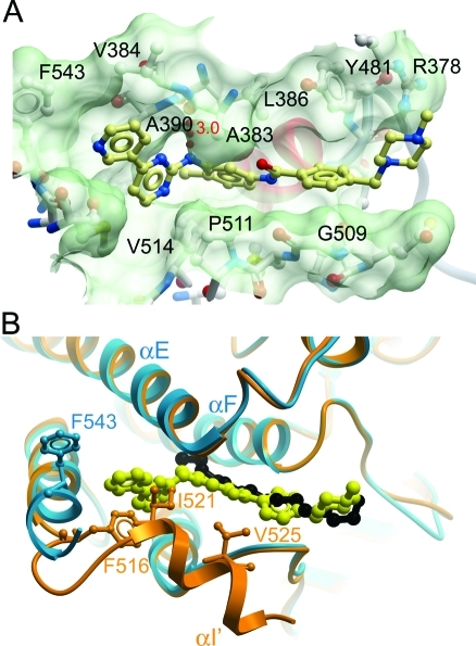
Myristate binding pocket of ABL2. (A) Surface of the myristate binding pocket of ABL2, with imatinib shown as a yellow ball-and-stick representation. J Med Chem. 2011 Apr 14;54(7):2359-67. |
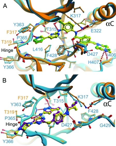
Comparison of ABL2:imatinib and ABL2:1 with ABL1:imatinib and ABL1:1. J Med Chem. 2011 Apr 14;54(7):2359-67. |
 m.cnreagent.com