| CAS NO: | 1032754-93-0 |
| 规格: | ≥98% |
| 包装 | 价格(元) |
| 5mg | 电议 |
| 25mg | 电议 |
| 50mg | 电议 |
| 100mg | 电议 |
| 250mg | 电议 |
| 500mg | 电议 |
Molecular Weight (MW) | 498.6 |
Formula | C23H30N8O3S |
CAS No. | 1032754-93-0 |
Storage | -20℃ for 3 years in powder form |
-80℃ for 2 years in solvent | |
Solubility (In vitro) | DMSO: 20 mg/mL (40.11 mM) |
Water:<1 mg/mL | |
Ethanol: <1 mg/mL | |
Solubility (In vivo) | 0.5% methylcellulose+0.2% Tween 80: 30 mg/mL |
Synonym | Apitolisib; GDC 0980; GDC-0980; GDC0980; RG7422; RG-7422; RG 7422; GNE 390; GNE-390; GNE390 |
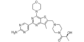
Chemical Name | (2S)-1-[4-[[2-(2-aminopyrimidin-5-yl)-7-methyl-4-morpholin-4-ylthieno[3,2-d]pyrimidin-6-yl]methyl]piperazin-1-yl]-2-hydroxypropan-1-one |
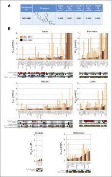
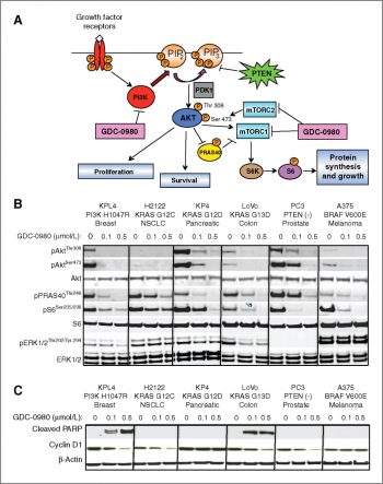
In Vitro | Kinase Assay: Enzymatic activity of the Class I PI3K isoforms is measured using a fluorescence polarization assay that monitors formation of the product 3,4,5-inositoltriphosphate molecule as it competes with fluorescently labeled PIP3 for binding to the GRP-1 pleckstrin homology domain protein. An increase in phosphatidyl inositide-3-phosphate product results in a decrease in fluorescence polarization signal as the labeled fluorophore is displaced from the GRP-1 protein binding site. Class I PI3K isoforms are expressed and purified as heterodimeric recombinant proteins. PI3K isoforms are assayed under initial rate conditions in the presence of 10 mM Tris (pH 7.5), 25 μM ATP, 9.75 μM PIP2, 5% glycerol, 4 mM MgCl2, 50 mM NaCl, 0.05% (v/v) Chaps, 1 mM dithiothreitol, 2% (v/v) DMSO at the following concentrations for each isoform: PI3Kα,β at 60 ng/mL; PI3Kγ at 8 ng/mL; PI3Kδ at 45 ng/mL. After assay for 30 minutes at 25°C, reactions are terminated with a final concentration of 9 mM EDTA, 4.5 nM TAMRA-PIP3, and 4.2 μg/mL GRP-1 detector protein before reading fluorescence polarization on an Envision plate reader. IC50s are calculated from the fit of the dose–response curves to a 4-parameter equation.Human recombinant mTOR(1360–2549) is expressed and purified from insect cells and assayed using a Lanthascreen fluorescence resonance energy transfer format in which phosphorylation of recombinant green fluorescent protein (GFP)-4-EBP1 is detected using a terbium-labeled antibody to phospho-threonine 37/46 of 4-EBP1. Reactions are initiated with ATP and conducted in the presence of 50 mM Hepes (pH 7.5), 0.25 nM mTOR, 400 nM GFP-4E-BP1, 8 μM ATP, 0.01% (v/v) Tween 20, 10 mM MnCl2, 1 mM EGTA, 1 mM dithiothreitol, and 1% (v/v) DMSO. Assays are conducted under initial rate conditions at room temperature for 30 minutes before terminating the reaction and detecting product in the presence of 2 nM Tb-anti-p4E-BP1 antibody and 10 mM EDTA. Dose–response curves are fit to an equation for competitive tight-binding inhibition and apparent Ki' s are calculated using the determined Km for ATP of 6.1 μM.
Cell Assay: Antiproliferative cellular assays are conducted using PC3 and MCF7.1 human tumor cell lines. MCF7.1 is an in vivo selected line and originally derived from the parental human MCF7 breast cancer cell line. Cell lines are cultured in RPMI supplemented with 10% fetal bovine serum, 100 units/mL penicillin, and 100 μg/mL streptomycin, 10 mM HEPES, and 2 mM glutamine at 3°C under 5% CO2. MCF7.1 cells or PC3 cells are seeded in 384-well plates in media at 1000 cells/well or 3000 cells/well, respectively, and incubated overnight prior to the addition of GDC-0980 to a final DMSO concentration of 0.5% v/v. MCF7.1 cells and PC3 cells are incubated for 3 days and 4 days, respectively, prior to the addition of CellTiter-Glo reagen and reading of luminescence using an Analyst plate reader. For antiproliferative assays, a cytostatic agent such as aphidicolin and a cytotoxic agent such as staurosporine are included as controls. Dose–response curves are fit to a 4-parameter equation and relative IC50s are calculated using Assay Explorer software.. GDC-0980 shows the potent and selective inhibitory activities against class I PI3K and mTOR kinase versus a large panel of kinases with Ki of 17 nM for mTOR and IC50 of 5 nM, 27 nM, 7 nM, and 14 nM for PI3Kα, β, δ, and γ, respectively. In vitro, GDC-0980 significantly inhibits cell proliferation in PC3 and MCF7 cells with IC50 of 307 nM and 255 nM, respectively. A recent study shows that GDC-0980 reduces cancer cell viability by inhibiting cell-cycle procession and inducing apoptosis with most potency in prostate (IC50 < 200 nM 50%),<500 nM 100%), breast (IC50 <200 nM 37%, <500 nM 78%) and NSCLC lines (IC50 <200 nM 29%, <500 nM 88%) and less potency in pancreatic (IC50 <200 nM 13%, <500 nM 67%) and melanoma cell lines (IC50 <200 nM 0%, <500 nM 33%). |
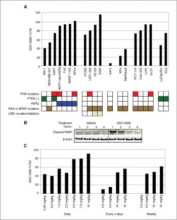
In Vivo | In both PC-3 and MCF-7 neo/HER2 xenograft models, GDC-0980 at a dose of 1 mg/kg, exhibits significant antitumor activity by causing tumor growth delay. Furthermore, GDC-0980 results in tumor stasis or regressions at the maximum tolerated dose of 7.5 mg/kg. In mice, intravenous GDC-0980 administration at 1 mg/kg leads to low clearance (Clp: 9.2 mL/min/kg, Vss: 1.7 L/kg). While, oral administration at 5 mg/kg in 80% PEG400 and at 50 mg/kg as a crystalline suspension in 0.5% methylcellulose/0.2% Tween-80 also results in favorable pharmacokinetic parameters. |
Animal model | PC3 and MCF7.1 cells are injected s.c. into the right hind flank of athymic nu/nu (nude) mice. |
Formulation & Dosage | Dissolved in 0.5% methylcellulose with 0.2% Tween-80 (MCT); 7.5 mg/kg; oral |
References | [1] Sutherlin DP, et al. J Med Chem, 2011, 54(21), 7579-7587. |
|
Mol Cancer Ther, 2011, 10(12), 2426-2436. |
|
 m.cnreagent.com