| CAS NO: | 1086062-66-9 |
| 规格: | ≥98% |
| 包装 | 价格(元) |
| 1mg | 电议 |
| 5mg | 电议 |
| 10mg | 电议 |
| 25mg | 电议 |
| 50mg | 电议 |
| 100mg | 电议 |
| 250mg | 电议 |
| 500mg | 电议 |
Molecular Weight (MW) | 505.5 |
Formula | C25H17F2N5O3S |
CAS No. | 1086062-66-9 |
Storage | -20℃ for 3 years in powder form |
-80℃ for 2 years in solvent | |
Solubility (In vitro) | DMSO: 100 mg/mL (197.8 mM) |
Water:<1 mg/mL | |
Ethanol: <1 mg/mL | |
Solubility (In vivo) | 1% DMSO+30% polyethylene glycol+1% Tween 80: 18 mg/mL |
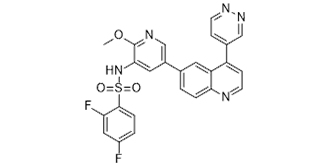
Synonym/Chemical Name | GSK2126458; GSK-2126458; GSK 2126458; GSK458; GSK 458; GSK-458; 2,4-difluoro-N-[2-methoxy-5-(4-pyridazin-4-ylquinolin-6-yl)pyridin-3-yl]benzenesulfonamide |
In Vitro | Kinase Assay: Compounds are serially diluted (3-fold in 100% DMSO) across a 384-well polypropylene mother plate from column 1 to column 12 and column 13 to column 24, to yield 11 concentrations for GSK2126458. Columns 6 and 18 contain only DMSO. Once titrations are made, 0.05μL is transferred to a 384-well low-volume assay plate. This assay plate contains three pharmacological controls (known PI3K inhibitors) and 3 assay controls: (1) Enzyme without inhibitor; (2) Buffer minus enzyme, and (3) Buffer minus enzyme plus native PIP3. DMSO is stamped into all wells of columns 6 and 18. PIP3 is added at 40 μM in 1X Reaction buffer (1μL of 200 μM PIP3) to alternating rows of column 18 (wells 18 B, D, F, H, J, L, N, P). The no-enzyme control reactions are run in wells 18 A, C, E, G, I, K, M, O (0.1μL of 100% DMSO). The PI3-Kinase profiling assay is optimized using the HTRF kit. The assay kit contains seven reagents: 1) 4X Reaction Buffer; 2) native PIP2 (substrate); 3) Stop A (EDTA); 4) Stop B (Biotin-PIP3); 5) Detection Mix A (Streptavidin-APC); 6) Detection Mix B (Eu-labeled Anti-GST plus GST-tagged PHdomain); 7) Detection Mix C (KF). PI3Kinase Reaction Buffer is prepared by diluting the stock 1:4 with de-ionized water. Freshly prepared DTT is added at a final concentration of 5 mM on the day of use. Enzyme addition and compound pre-incubation are initiated by the addition of 2.5μL of PI3K (at twice its final concentration) in 1X reaction buffer to all wells using a Multidrop Combi. Plates are incubated at room temperature for 15 minutes. Reactions are initiated by addition of 2.5μL of 2X substrate solution (PIP2 and ATP in 1X reaction buffer) using a Multidrop Combi. Plates are incubated at room temperature for one hour. Reactions are quenched by the addition of 2.5μL of stop solution (Stop A and Stop B pre-mixed at a ratio of 5:1, respectively) to all wells using the Multidrop Combi. The quenched reactions are then processed to detect product formation by adding 2.5μL of Detection Solution to all wells using the Mulitdrop Combi (Detection mix C, Detection mix A, and Detection mix B combined together in an 18:1:1 ratio, i.e.: for a 6000 μL total volume, mix 5400 μL Detection mix C, 300μL Detection mix A, and 300 μL Detection mix B. Note: this solution should be prepared 2 hours prior to use). Following a one hour incubation in the dark, the HTRF signal is measured on the Envision plate reader set for 330nm excitation and dual emission detection at 620nm (Eu) and 665nm (APC).
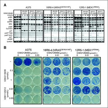
Cell Assay: BT474, HCC1954 and T-47D (human breast) are cultured in RPMI-1640 containing 10% fetal bovine serum at 37 °C in 5% CO2 incubator. Cells are split into T75 flask two to three days prior to assay set up at density which yields approximately 70-80% confluence at time of harvest for assay. Cells are harvested using 0.25% trypsin-EDTA. Cell counts are performed on cell suspension using Trypan Blue exclusion staining. Cells are then plated in 384 well black flat bottom polystyrene in 48 μL of culture media per well at 1,000 cells/well. All plates are placed at 5% CO2, 37 °C overnight and GSK2126458 is added the following day. One plate is treated with CellTiter-Glo for a day 0 (t=0) measurement and read as described below. GSK2126458 is prepared in clear bottom polypropylene 384 well plates with consecutive two fold dilutions. 4 μL of these dilutions are added to 105 μL culture media, after mixing the solution, 2 μL of these dilutions are added into each well of the cell plates. The final concentration of DMSO in all wells is 0.15%. Cells are incubated at 37 °C, 5% CO2 for 72 hours. Following 72 hours of incubation with GSK2126458 each plate is developed and read. CellTiter-Glo reagent is added to assay plates using a volume equivalent to the cell culture volume in the wells. Plates are shaken for approximately two minutes and incubated at room temperature for approximately 30 minutes and chemiluminescent signal is read on the Analyst GT reader. Results are expressed as a percent of the t=0 and plotted against the GSK2126458 concentration. Cell growth inhibition is determined for GSK2126458 by fitting the dose response with a 4 or 6 parameter curve fit using XLfit software and determining the concentration that inhibits 50% of the cell growth (gIC50) with the Y min as the t=0 and Y max as the DMSO control. Value from wells with no cells is subtracted from all samples for background correction. GSK2126458 potently inhibits the activity of common activating mutants of p110α (E542K, E545K, and H1047R) found in human cancer with Ki of 8 pM, 8 pM and 9 pM, respectively. GSK2126458 causes a significant reduction in the levels of pAkt-S473 with remarkable potency in T47D and BT474 cells with IC50 of 0.41 nM and 0.18 nM, respectively. Furthermore, GSK2126458 leads to a G1 cell cycle arrest and produces the inhibitory effect on cell proliferation in a large panel of cell lines, including T47D and BT474 breast cancer lines with IC50 of 3 nM and 2.4 nM, respectively. |
In Vivo | In a BT474 human tumor xenograft model, GSK2126458 treatment results in a dose-dependent reduction in pAkt-S473 levels, and exhibited dose-dependent tumor growth inhibition at a low dose of 300 μg /kg. Besides, GSK2126458 shows low blood clearance and good oral bioavailability in four preclinical species (mouse, rat, dog, and monkey). |
Animal model | Human BT474 tumors implanted in mice. |
Formulation & Dosage | Dissolved in DMSO and then diluted in water; ≤300 μg /kg; p.o. |
References | [1] Knight SD, et al. ACS Med. Chem. Lett. 2010, 1 (1), 39–43.; [2] Greger JG, et al. Mol Cancer Ther. 2012, 11(4), 909-920. |
Greger JG, et al. Mol Cancer Ther. 2012, 11(4), 909-920. |
Developing selective type II kinase inhibitors. (A) Docking imatinib into the X-ray co-crystal structure of DDR1. (B) Chemical structure of DDR1-IN-1/2 and representative developing rationale. ACS Chem Biol. 2013 Oct 18;8(10):2145-50. |
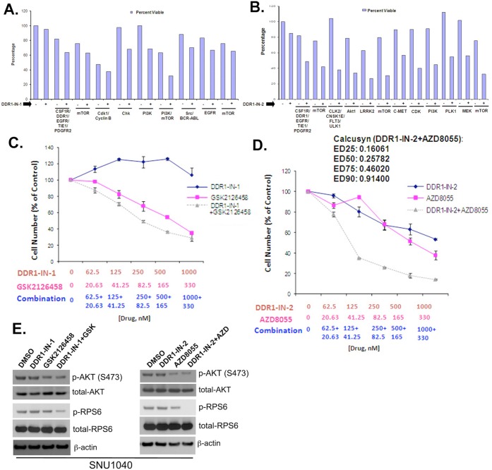
Combinatorial Screening of DDR1-IN-1/2 with the LINCS library against the SNU-1040 cell line. ACS Chem Biol. 2013 Oct 18;8(10):2145-50. |
 m.cnreagent.com