| CAS NO: | 393105-53-8 |
| 规格: | ≥98% |
| 包装 | 价格(元) |
| 5mg | 电议 |
| 10mg | 电议 |
| 25mg | 电议 |
| 50mg | 电议 |
| 100mg | 电议 |
| 250mg | 电议 |
| 500mg | 电议 |
| Molecular Weight (MW) | 439.38 |
|---|---|
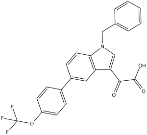
| Formula | C24H16F3NO4 |
| CAS No. | 393105-53-8 |
| Storage | -20℃ for 3 years in powder form |
| -80℃ for 2 years in solvent | |
| Solubility (In vitro) | DMSO: 72 mg/mL (163.9 mM) |
| Water: <1 mg/mL | |
| Ethanol: 18 mg/mL (41 mM) | |
| Solubility (In vivo) | 2.0% Tween 80+0.5% methylcellulose: 30mg/mL |
| Synonyms | PAI039; PAI-039; PAI 039. |
| In Vitro | In vitro activity: In a panel of human bladder cell lines, PAI-1 results in the reduction of cellular proliferation, cell adhesion, and colony formation, and the induction of apoptosis and anoikis. Kinase Assay: The chromogenic assay is initiated by the addition of tiplaxtinin (10 – 100 μM final concentration, maximum DMSO concentration of 0.2%) to recombinant human PAI-1 (140 nM in pH 6.6 buffer). After a 15 minute incubation at 25°C, 70 nM of recombinant human t-PA is added, and the combination of tiplaxtinin, PAI-1 and tPA are incubated for an additional 30 minutes. After the second incubation, Spectrozyme tPA, is added and absorbance read at 405 nm at 0 and 60 minutes. Relative PAI-1 inhibitory activity is equal to the residual tPA activity in the tiplaxtinin / PAI-1 treatment. Control treatments include the complete inhibition of tPA by PAI-1 at the molar ratio employed (2:1), and the absence of any effect of the tiplaxtinin on t-PA alone. The immunofunctional assay is based upon the non-SDS dissociable interaction between tPA and active PAI-1. Assay plates are coated with 100 μl of a solution of t-PA (10 μg/ml in TBS), and kept at 4 °C overnight. Tiplaxtinin is dissolved in DMSO and diluted to a final concentration of 1-100 μM as described above. Tiplaxtinin is then incubated with human PAI-1 (50 ng/ml) for 15 minutes, and an aliquot of this solution added to the t-PA-coated plate for 1 h. The solution is aspirated from the plate, which is then washed with a buffer consisting of 0.05% Tween 20 and 0.1% BSA in TBS. This assay detects only active inhibitory PAI-1 (not latent or substrate) bound to the plate, and is quantitated using a monoclonal antibody against human PAI-1 (MA33B8). A 1000X dilution of MA33B8 is added to the plate and incubated at for one hour, aspirated and washed. A secondary antibody consisting of goat anti-mouse IgG (H+L)-AP alkaline phosphatase conjugate is added, incubated for one hour, aspirated and washed. A 100 μl aliquot of alkaline phosphatase solution is added, followed by determination of absorbance at 405 nm 60 minutes later. The quantitation of residual active PAI-1 bound to t-PA at varying concentrations of tiplaxtinin is used to determine the IC50 by fitting the results to a logistic dose-response program, with the IC50 defined as the concentration of compound required to achieve 50% inhibition of PAI-1 activity. The assay sensitivity is 5 ng/ml of human PAI-1 as determined from a standard curve ranging from 0-100 ng/ml of human PAI-1. Cell Assay: Briefly, cell lines, T24, UM-UC-14, UROtsa, and HeLa cells are plated in 96-well dishes in triplicate at 1 ?103 cells per well and allowed to adhere for 24 hours. Subsequently, tiplaxtinin is added to the wells and allowed to incubate at the indicated concentrations. Cellular proliferation is determined by CellTiter-Glo Luminescent Cell Viability Assay according to manufacturer's instructions at 24 hours, and IC50 of tiplaxtinin is determined in Graphpad Prism. Luminescence was measured using a FLUOstar OPTIMA Reader. |
|---|---|
| In Vivo | In a rat carotid thrombosis model, Tiplaxtinin (1 mg/kg, p.o.) increases time to occlusion and prevents the carotid blood flow reduction. In C57BL/6J mice, (1 mg/g chow) attenuates Ang II-induced aortic remodeling. In untreated type 1 diabetic mice, Tiplaxtinin (p.o.) restores skeletal muscle regeneration. In athymic mice bearing human cancer cell line T24 and HeLa xenografts, Tiplaxtinin (1 mg/kg, p.o.) reduces tumor xenograft growth, associated with a reduction in tumor angiogenesis, a reduction in cellular proliferation, and an increase in apoptosis. |
| Animal model | Rat with carotid thrombosis |
| Formulation & Dosage | Formulated in 2.0% Tween 80/0.5% methylcellulose; 1 mg/kg; p.o. |
| References | J Med Chem. 2004 Jul 1;47(14):3491-4; Arterioscler Thromb Vasc Biol. 2005 Feb;25(2):365-71; Mol Cancer Ther. 2013 Dec;12(12):2697-708. |
 m.cnreagent.com