| CAS NO: | 143851-98-3 |
| 规格: | ≥98% |
| 包装 | 价格(元) |
| 5mg | 电议 |
| 10mg | 电议 |
| 25mg | 电议 |
| 50mg | 电议 |
| 100mg | 电议 |
| 250mg | 电议 |
| 500mg | 电议 |
| Molecular Weight (MW) | 600.1 |
|---|---|
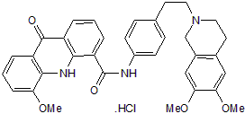
| Formula | C34H33N3O5.HCl |
| CAS No. | 143851-98-3 (HCl); |
| Storage | -20℃ for 3 years in powder form |
| -80℃ for 2 years in solvent | |
| Solubility (In vitro) | DMSO: 10 mM |
| Water: N/A | |
| Ethanol: N/A | |
| Solubility (In vivo) | O=C1C2=CC=CC(C(NC(C=C3)=CC=C3CCN4CC(C=C(C(OC)=C5)OC)=C5CC4)=O)=C2NC6=C1C=CC=C6OC |
| Synonyms | GW120918; GF-120918; GW120918; GW 120918; GW-0918;GW0918; GW 0918; GG-918;GG918;GG 918; |
| In Vitro | In vitro activity: Elacridar inhibits P-glycoprotein (P-gp) labeling by [3H]azidopine with a IC50 of 0.16 μM. In Caki-1 and ACHN cells, elacridar ( 2.5 μM) significantly ihibits the cell growth. The P-glycoprotein activity is found to be inhibited by elacridar. The combination of elacridar and sunitinib lead to a significant reduction in ABC Sub-family B Member 2 (ABCG2) expression in 786-O cells. Kinase Assay: 10 μL of unlabeled cell membrane suspension (at 0.4 mg of protein/mL) are aliquoted into each well in 96-well plates. 5 μL of GF120918 are then added to each well. The plate is incubated 25 min at 25℃ in the dark. 5 μL of tritiated azidopine (1.8 TBq/mmol) (0.6 μM in HCI 0.2 mM) are added to each well. After 25 min of incubation at 25℃ in the dark, samples are simultaneously irradiated for 2 min at 254 nm at 0℃ with a thin layer chromatography-designed UV lamp directly in contact with the plate. Samples are solubilized in sodium dodecyl sulfate-polyacrylamide gel electrophoresis sample buffer but not heated. After separation on a 7.5% polyacrylamide gel, the gel is treated for fluorography with Amplify and exposed during 3 days onto a photosensitive film. The fluorography is analysed using a Camag thin layer chromatography Scanner II densitometer. Cell Assay: 3.0×10³ cells per well are seeded in a 96-well plate. After 24 h incubation, an optimum concentration gradient of elacridar is added to each well. After culturing for 48 h, cell viability is assessed using the proliferation reagent, MTT. Control cells are treated with the vehicle only, 0.1% DMSO. After this final incubation, the medium is aspirated and precipitated formazan crystals are dissolved in DMSO (100 μL/well). The absorbance of each well is measured at 540 nm, and a reference wavelength of 650 nm is read with a multiskan JX microplate reader. Cell viability is calculated as percentage of the control value. |
|---|---|
| In Vivo | Oral co-administration of elacridar (100 mg/kg, p.o.) and crizotinib increases the plasma and brain concentrations and brain-to-plasma ratios of crizotinib in wild-type mice, equaling the levels in Abcb1a/1b; Abcg2-/- mice. In friend leukemia virus stain B mice, the brain-to-plasm partition coefficient (Kp, brain) of elacridar is 0.82, 0.43, and 4.31 after intravenous (2.5 mg/kg), intraperitoneal (100 mg/kg), and oral (100 mg/kg) treatment, respectively. In Mrp4(-/-) mice, elacridar fully inhibits P-gp mediated transport of topotecan, without siginificant effects on Bcrp1-mediated transport. |
| Animal model | Male wild-type, Abcb1a/1b-/-34, Abcg2 -/-32 and Abcb1a/1b;Abcg2 |
| Formulation & Dosage | Dissolved in mixture of DMSO, Polysorbate 80, ethanol, and water (1.8:1.17:6.03:1, v/v/v/v); 100 mg/kg ; p.o. |
| References | Int J Cancer. 2014 Mar 15;134(6):1484-94; Drug Metab Dispos. 2012 Aug;40(8):1612-9; Clin Cancer Res. 2007 Nov 1;13(21):6440-9. |
 m.cnreagent.com