| CAS NO: | 1052532-15-6 |
| 规格: | ≥98% |
| 包装 | 价格(元) |
| 5mg | 电议 |
| 10mg | 电议 |
| 25mg | 电议 |
| 50mg | 电议 |
| 100mg | 电议 |
| 250mg | 电议 |
| 500mg | 电议 |
| Molecular Weight (MW) | 479.4 |
|---|---|
| Formula | C24H28Cl2N2O4 |
| CAS No. | 1052532-15-6 |
| Storage | -20℃ for 3 years in powder form |
| -80℃ for 2 years in solvent | |
| Solubility (In vitro) | DMSO: 95 mg/mL (198.2 mM) |
| Water:<1 mg/mL | |
| Ethanol: <1 mg/mL | |
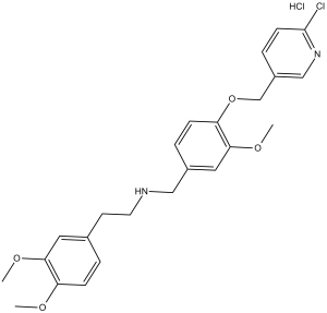
| Other info | Chemical Name: N-(4-((6-chloropyridin-3-yl)methoxy)-3-methoxybenzyl)-2-(3,4-dimethoxyphenyl)ethanamine hydrochloride InChi Key: QBGSVDJLQQXEGG-UHFFFAOYSA-N InChi Code: InChI=1S/C24H27ClN2O4.ClH/c1-28-20-7-4-17(12-22(20)29-2)10-11-26-14-18-5-8-21(23(13-18)30-3)31-16-19-6-9-24(25)27-15-19;/h4-9,12-13,15,26H,10-11,14,16H2,1-3H3;1H SMILES Code: COC1=CC=C(CCNCC2=CC=C(OCC3=CC=C(Cl)N=C3)C(OC)=C2)C=C1OC.[H]Cl |
| Synonyms | SBE13 HCl; SBE 13; SBE13; SBE-13 HCl |
| In Vitro | In vitro activity: SBE 13 decreases cell proliferation in various cancer cell lines, and causes a G2/M arrest followed by apoptosis. In primary cells, SBE 13 does not impair cell cycle and thus proliferation of primary cells. SBE13 in combination with Enzastaurin displays a synergistic reduction of cell proliferation and enhanced apoptosis induction in HCT116(p53-/-) cells. Kinase Assay: To assay Plk1 and Aurora A kinase activity, cells are lysed after 13 hrs release in the presence of SBE13 after double thymidine block, and kinases are immunoprecipitated from lysates using antibodies as described. In brief, for each immunoprecipitation 800 μg of total protein were incubated with 1.5 μg Plk1 antibody cocktail, 3 μg Plk2 antibody, 3 μg Plk3 antibody, or 5 μg Aurora A antibody, respectively, for 2 hrs at 4°C on a rotator. Immunoprecipitated protein is collected using Protein G Agarose beads. The Plk1, Plk2 and Plk3 immunoprecipitates are incubated with 1 μg casein and with 1 μCi of [γ32-P]ATP for 30 min at 37°C in kinase buffer. The Aurora A immunoprecipitates are incubated with 0.5 μl Histone and with 1 μCi of [γ32-P]ATP for 60 min at room temperature in kinase buffer. Products from the kinase assays are fractionated on 10% Bis-Tris-polyacrylamide gels, and the phosphorylated substrate is visualized by autoradiography after an exposure of 12 to 36 hrs. An equal amount of immunoprecipitates is subjected to western blot analysis to confirm equal loading of Plk1, Plk2, Plk3 or Aurora A protein in kinase reactions. Immunoprecipitated Plk1 after 13 hrs release in the presence of SBE13 is assayed after de-phosphorylation using λ protein phosphatase and compared to kinase activity of endogenous immunoprecipitated Plk1. Activity of Plk1 kinase with and wiiiuithout de-phosphorylation is compared and the ratio between de-phosphorylated and “normal” endogenous immunoprecipitated Plk1 kinase activity is calculated. Cell Assay: Cells (HeLa, A431, HCT-15, HT-29, MCF-7, U2OS, LN-229, SKW 6.4, PC-3, Det-562, SK-BR-3, SK-OV-3 and A549nb cells) are treated with SBE13 one day after subculturing. Control cells are incubated with normal culture medium. Concentrations of SBE13 ranged from 1 nM–100 μM. The growth rate of 1 x 10⁵ cells per 6-well is determined by counting cells at 24, 48 and 72 hours after treatment. Cell culture studies are performed in triplicate for each time point. |
|---|---|
| In Vivo | |
| Animal model | |
| Formulation & Dosage | |
| References | Cell Cycle. 2010 Feb 15;9(4):761-73; Oncotarget. 2014 Apr 30;5(8):2263-75. |
 m.cnreagent.com