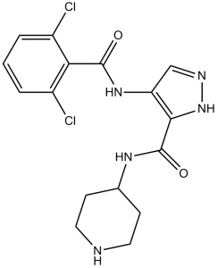
| CAS NO: | 844442-38-2 |
| 规格: | ≥98% |
| 包装 | 价格(元) |
| 5mg | 电议 |
| 10mg | 电议 |
| 25mg | 电议 |
| 50mg | 电议 |
| 100mg | 电议 |
| 250mg | 电议 |
| 500mg | 电议 |
| Molecular Weight (MW) | 382.24 |
|---|---|
| Formula | C16H17Cl2N5O2 |
| CAS No. | 844442-38-2 |
| Storage | -20℃ for 3 years in powder form |
| -80℃ for 2 years in solvent | |
| Solubility (In vitro) | DMSO: 10 mg/mL (26.1 mM) |
| Water: <1 mg/mL | |
| Ethanol:<1 mg/mL | |
| Solubility (In vivo) | 30% Propylene glycol, 5% Tween 80, 65% D5W: 30 mg/mL |
| Synonyms | AT-7519; AT7519; AT 7519 |
| In Vitro | In vitro activity: AT7519 is an ATP competitive CDK inhibitor with a Ki value of 38 nM for CDK1. AT7519 is inactive against all non-CDK kinases with the exception of GSK3β (IC50 = 89 nM). AT7519 shows potent antiproliferative activity in a variety of human tumor cell lines with IC50 values ranging from 40 nM for MCF-7 to 940 nM for SW620 consistent with the inhibition of CDK1 and CDK2. AT7519 induces dose-dependent cytotoxicity in multiple myeloma (MM) cell lines with IC50 values ranging from 0.5 to 2 μM at 48 hours, with the most sensitive cell lines being MM.1S (0.5 μM) and U266 (0.5 μM) and the most resistant MM.1R (>2 μM). It does not induce cytotoxicity in peripheral blood mononuclear cells (PBMNC). AT7519 partially overcomes the proliferative advantage conferred by IL6 and IGF-1 as well as the protective effect of bone marrow stromal cells (BMSCs). AT7519 induces rapid dephosphorylation of RNA pol II CTD at serine 2 and serine 5 sites, and leads to the inhibition of transcription, partially contributing to AT7519 induced cytotoxicity of MM cells. AT7519 induces activation of GSK-3β by down-regulating GSK-3β phosphorylation, which also contributes to AT7519 induced apoptosis independent of the inhibition of transcription. Kinase Assay: Kinase assays for CDK1, CDK2 and GSK3-β are all carried out in a radiometric filter binding format. Assays for CDK5 are in DELFIA format and for CDKs 4 and 6 in ELISA format. For CDKs 1 and 2, the relevant CDK and 0.12 μg/mL Histone H1 are incubated in 20 mM MOPS, pH 7.2, 25 mM β-glycerophosphate, 5 mM EDTA, 15 mM MgCl2, 1 mM sodium orthovanadate, 1 mM DTT, 0.1 mg/mL BSA, 45 μM ATP (0.78 Ci/mmol) and different concentrations of AT7519 for 2 or 4 hours respectively. For GSK3-β, the relevant enzyme and 5 μM glycogen synthase peptide 2 along with 10 mM MOPS pH 7.0, 0.1 mg/mL BSA, 0.001% Brij-35, 0.5% glycerol, 0.2 mM EDTA, 10 mM MgCl2, 0.01% β-mercaptoethanol, 15 μM ATP (2.31 Ci/mmol) and different concentrations of AT7519 are incubated for 3 hours. Assay reactions are stopped by adding an excess of orthophosphoric acid and filtered using Millipore MAPH filter plates. The plates are then washed, scintillant added and radioactivity measured by scintillation counting on a Packard TopCount. For CDK5, CDK5/p35 and 1μM of a biotinylated Histone H1 peptide (Biotin-PKTPKKAKKL) are incubated in 25 mM Tris-HCl, pH 7.5, 2.5 mM MgCl2, 0.025% Brij-35, 0.1 mg/mL BSA, 1 mM DTT, 15 μM ATP and different concentrations of AT7519 for 30 minutes. Assay reactions are stopped using EDTA, transferred to Neutravidin-coated plates and phosphorylated peptide quantified by means of a rabbit phospho-cdk1 substrate polyclonal antibody and DELFIA europium-labelled anti-rabbit IgG secondary antibody using time-resolved fluorescence at λex=335nm, λem=620nm. For CDK 4 and 6 assays, plates are coated with GST- pRb769-921 and blocked with Superblock. CDK4 or 6 is incubated with 15 mM MgCl2, 50 mM HEPES, pH 7.4, 1 mM DTT, 1 mM EGTA, pH 8.0, 0.02% Triton X-100, 2.5% DMSO and different concentrations of AT7519; the reaction is initiated by addition of ATP. After 30 minutes, reactions are stopped by the addition of 0.5 M EDTA pH 8.0. Plates are then washed and incubated for one hour with the primary antibody (anti- p-Rb Serine 780) diluted in Superblock followed by secondary antibody (alkaline phosphatase linked anti-rabbit) for a further hour. Plates are developed using the Attophos system and fluorescence read on a Spectramax Gemini plate reader at excitation 450 nm and emission 580 nm. In all cases, IC50 values are calculated from replicate curves, using GraphPad Prism software. Cell Assay: Cells (MM.1S, MM.1R, RPMI8226, U266, RPMI8266, RPMI-Dox40, OPM1 cells, primary MM cells and PBMNCs) are incubated with different concentrations of AT7519 for 24 or 48 hours at 37 °C. Cell viability is assessed by measuring 3-(4,5-dimethylthiazol-2-yl)-2,5 diphenyl tetrasodium bromide (MTT) dye absorbance. DNA synthesis is measured by tritiated thymidine uptake (3H-TdR). Apoptosis is assessed by using Annexin V/PI staining. The percentage of cells undergoing apoptosis is defined as the sum of early apoptosis (Annexin V-positive cells) and late apoptosis (Annexin V-positive and PI-positive cells. |
|---|---|
| In Vivo | A twice daily dosing of AT7519 (9.1 mg/kg) causes tumor regression of both early-stage and advanced-stage s.c. tumors in the HCT116 and HT29 colon cancer xenograft models. AT7519 treatment (15 mg/kg) inhibits tumor growth and prolongs the median overall survival of mice in the human MM xenograft mouse model in association with increased caspase 3 activation. |
| Animal model | Male SCID mice inoculated subcutaneously with MM.1S cells |
| Formulation & Dosage | Dissolved in 0.9% saline; 15 mg/kg/day; i.p. injection |
| References | Mol Cancer Ther. 2009 Feb;8(2):324-32; Oncogene. 2010 Apr 22;29(16):2325-36. |
 m.cnreagent.com