| CAS NO: | 186692-46-6 |
| 规格: | ≥98% |
| 包装 | 价格(元) |
| 10mg | 电议 |
| 25mg | 电议 |
| 50mg | 电议 |
| 100mg | 电议 |
| 250mg | 电议 |
| 500mg | 电议 |
| 1g | 电议 |
| Molecular Weight (MW) | 354.45 |
|---|---|
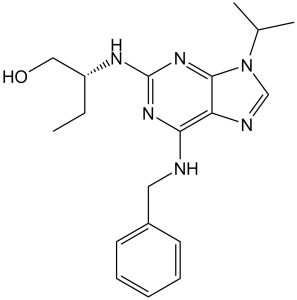
| Formula | C19H26N6O |
| CAS No. | 186692-46-6 |
| Storage | -20℃ for 3 years in powder form |
| -80℃ for 2 years in solvent | |
| Solubility (In vitro) | DMSO: 71 mg/mL (200.3 mM) |
| Water: <1 mg/mL | |
| Ethanol: 6 mg/mL (16.9 mM) | |
| Solubility (In vivo) | 1% DMSO+30% polyethylene glycol+1% Tween 80: 30 mg/mL |
| Synonyms | Seliciclib, R-Roscovitine; CYC202; CYC202; Roscovitin; Roscovitine; CYC202; CYC 202; Chemical Name: (R)-2-((6-(benzylamino)-9-isopropyl-9H-purin-2-yl)amino)butan-1-ol SMILES Code: CC[C@@H](NC1=NC(NCC2=CC=CC=C2)=C3N=CN(C(C)C)C3=N1)CO Exact Mass: 354.21681 |
| In Vitro | In vitro activity: Roscovitine displays high efficiency and high selectivity towards some cyclin-dependent kinases with IC50 of 0.65, 0.7, 0.7 and 0.16 μM for cdc2/cyclin B, cdk2/cyclin A, cdk2/cyclin E and cdk5/p53, respectively. Roscovitine reversibly inhibits the prophaselmetaphase transition in the micromolar range of starfish oocytes and sea urchin embryos, inhibits in vitro M-phase-promoting factor activity and in vitro DNA synthesis in Xenopus egg extracts, and suppresses the proliferation of mammalian cell lines with an average IC50 of 16 μM. In mesangial cells, Roscovitine results in a dose-dependent reduction of CDK2 activity that at concentrations of 7.5, 12.5 and 25 mM, Roscovitine causes a 25, 50% and 100% decrease in CDK2 activity, respectively. A recent study shows that Roscovitine inhibits cdk5 kinase activity, cell proliferation, multicellular development, and cdk5 nuclear translocation in Dictyostelium discoideum, without affecting the expression of cdk5 protein during axenic growth. Kinase Assay: Kinases activities are assayed at 30 °C in buffer C. Blank values are subtracted from the data and activities calculated as molar amount of phosphate incorporated in protein acceptor during a 10-minute incubation. Controls are performed with appropriate dilutions of DMSO. In a few cases, phosphorylation of the substrate is assessed by autoradiography after SDS/PAGE. p34cdc2/cyclin B is purified from M-phase starfish (M. glacialis) oocytes by affinity chromatography. It is assayed with 1 mg histone Hl/mL, in the presence of 15 μM [γ-32P]ATP (3000 Ci/mmol; 1 mCi/mL) in a final volume of 30 μL. After a 10-minute incubation at 30 °C, 25-μL aliquots of supernatant are spotted onto pieces of Whatman P81 phosphocellulose paper, and, after 20 seconds, the filters are washed five times (for at least 5 minutes each time) in a solution of 10mL phosphoric acid/L water. The wet filters are transferred into 6-mL plastic scintillation vials, 5 mL ACS scintillation fluid is added and the radioactivity measured in a Packard counter. The kinase activity is expressed as molar amount of phosphate incorporated in histone H1 during a 10-minutes incubation or as a percentage of maximal activity. p33cdk2/cyclin A and p33cdk2/cyclinE are reconstituted from extracts of sf9 insect cells infected with various baculoviruses. Cyclins A and E are fusion proteins with glutathione S-transferase and the complexes are purified on glutathione-agarose beads. Kinase activities are assayed with 1 mg/mL histone H1, in the presence of 15 μM [γ-32P]ATP, during 10 minutes, in a final volume of 30 μL, as described for the p34cdc2/cyclin B kinase. p33cdk5/p35 is purified from bovine brain, excluding the Mono S-chromatographic step. The active fractions from the Superose 12 column are pooled and concentrated to a final concentration of approximately 25 μg enzyme/mL. The kinase is assayed with 1 mg/mL histone HI in the presence of 15 μM [γ-32P]ATP, during 10 minutes in a final volume of 30 μL, as described for the p34cdc2/cyclin B kinase. p33cdk5/cyclin D1 is obtained from insect cell lysates. Cdk4 is a fusion protein with glutathione-S-transferase and the active complex is purified on glutathione-agarose beads. Its kinase activity is assayed with purified retinoblastoma protein (complexed with glutathione-S-transferase) in the presence of 15 μM [γ-32P]ATP, in a final volume of 30 μL. After a 15-minute incubation, 30 μL Laemmli sample buffer is added. The phosphorylated substrate is resolved by 10 % SDS/PAGE and analysed by autoradiography by overnight exposure to Hyperfilm MP and densitometry. p33cdk4/cyclinD 2 is obtained from insect cell lysates. It is assayed with purified retinoblastoma protein (complexed with glutathione-S-transferase) in the presence of 15 μM [γ-32P]ATP in a final volume of 30 μL. After a 30-minute incubation, 30 μL Laemmli sample buffer is added. The phosphorylated substrate is resolved by 10% SDS/PAGE and analysed by autoradiography by overnight exposure to Hyperfilm MP and densitometry. MAP kinase erkl (tagged with glutathione-S-transferase), is expressed in bacteria, purified on glutathione-agarose beads and assayed with 1 mg myelin basic protein/ml in the presence of 15 μM [γ-32P]ATP as described above for the p34cdc2cyclin B kinase. His-tagged erkl and erk2 are activated in vitro by mitogen-activated protein kinase kinase, purified (Ni-affinity and Mono Q) and assayed as described above during 10 minutes in a final volume of 30 μL. Protein kinase C isoforms are purified from baculovirus infected sf9 insect cells and assayed with 1 mg/mL protamine sulfate in the presence of 15 μM [γ-32P]ATP, during 10 minutes at 30 °C, in a final volume of 30 μL. Phosphorylated protamine sulfate is recovered on Whatman P81 phosphocellulose paper as described for the cdc2 kinase. The catalytic subunit of cAMP-dependent protein kinase, purified from bovine heart, is assayed with 1 mg histone Hl/ml, in the presence of 15 μM [γ-32P]ATP as described for the p34cdc2/cyclin B kinase. cGMP-dependent protein kinase, purified to homogeneity from bovine tracheal smooth muscle, is assayed with 1 mg histone Hl/mL, in the presence of 15 μM [γ-32P]ATP as described for the p34cdc2/cyclin B kinase. Casein kinase 2 is isolated from rat liver cytosol and assayed with 1 mg casein/mL and 15 μM [γ-32P]ATP. The substrate is spotted on Whatmann 3MM filters and washed with 10% (mass/vol.) trichloroacetic acid. Myosin light chain kinase, purified from chicken gizzard is assayed in the presence of 100 nM calmodulin, 100 μM CaCl2, 50 mM Hepes, 5 mM MgCI,, 1 mM dithiothreitol and 0.1 mg BSA/ml at pH 7.5 using a synthetic peptide based on the smooth-muscle myosin light-chain phosphorylation site and in the presence of 15 μM [γ-32P]ATP, in a final volume of 50 μL. Incorporation of radioactive phosphate is monitored on phosphocellulose filters as described above. ASK-γ, a plant homologue of GSK-3, is expressed as a glutathione-S-transferase fusion protein in Escherichia coli and purified on glutathione-agarose. ASK-γ kinase is assayed, for 10 minutes at 30 °C, with 5 μg myelin basic protein, in the presence of 15 μM [γ-32P]ATP in a final volume of 30 μL. The phosphorylated myelin basic protein is recovered on Whatman P81 phosphocellulose paper as described for the p34cdc2/cyclin B kinase. Insulin receptor tyrosine kinase domain (CIRK-41) is overexpressed in a baculovirus system and purified to homogeneity. Its kinase activity is assayed, for 10 minutes at 30 °C, with 5 μg Raytide, in the presence of 15 μM [γ-32P]ATP, in a final volume of 30 μL. The phosphorylated Raytide is recovered on Whatman P81 phosphocellulose paper as described for the p34cdc2/cyclin B kinase. c-src kinase is purified from infected Sf9 cells. The v-abl kinase is expressed in E. coli and affinity purified on IgG Affigel 10. Both kinases are assayed for 10 minutes at 30 °C, with 5 μg Raytide, in the presence of 15 μM [γ-32P]ATP, in a final volume of 30 μL. The phosphorylated Raytide is recovered on Whatman P81 phosphocellulose paper as described for the p34cdc2/cyclin B kinase. Cell Assay: 60 human tumour cell lines (Leukemia, non-small cell lung cancer, colon cancer, central nervous system cancer, melanoma, ovarian cancer, renal cancer, prostate cancer, breast cancer) comprising nine tumor types are cultured for 24 hours prior to a 48-hour continuous exposure to 0.01-100 μM roscovitine. A sulforhodaminine B protein assay is used to estimate the cytotoxicity. |
|---|---|
| In Vivo | Roscovitine, at a dose of 50 mg/kg, significantly inhibits growth of The Ewing's sarcoma family of tumors (ESFT) xenografts. Roscovitine enhances the antitumor effect of doxorubicin without increased toxicity with a mechanism that involves cell cycle arrest rather than apoptosis in nude mice bearing established MCF7 xenografts. |
| Animal model | A4573 cells are injected s.c. into the right posterior flank of CD1 nu/nu mice |
| Formulation & Dosage | Dissolved in either absolute methanol or DMSO and then diluted in 10% Tween 80, 20% N-N-dimethylacetamide, and 70% polyethylene glycol 400; ≤50 mg/kg; i.p. injection |
| References | Eur J Biochem. 1997 Jan 15;243(1-2):527-36; Cancer Res. 2005 Oct 15;65(20):9320-7. |
 m.cnreagent.com