| CAS NO: | 887375-67-9 |
| 规格: | ≥98% |
| 包装 | 价格(元) |
| 5mg | 电议 |
| 10mg | 电议 |
| 25mg | 电议 |
| 50mg | 电议 |
| 100mg | 电议 |
| 250mg | 电议 |
| 500mg | 电议 |
| Molecular Weight (MW) | 395.88 |
|---|---|
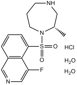
| Formula | C15H18FN3O2S. HCl. 2 H2O |
| CAS No. | 887375-67-9 |
| Storage | -20℃ for 3 years in powder form |
| -80℃ for 2 years in solvent | |
| Solubility (In vitro) | DMSO: 26 mg/mL warmed (65.7 mM) |
| Water: 79 mg/mL (199.5 mM) | |
| Ethanol: 5 mg/mL (12.6 mM) | |
| SMILES | FC1=CN=CC2=C1C(S(N3CCCNC[C@@H]3C)(=O)=O)=CC=C2.[H]Cl.O.O |
| Synonyms | K-115; trade name: Glanatec; K115; K 155; Ripasudil |
| In Vitro | In vitro activity: In monkey trabecular meshwork (TM) cells, Ripasudil induces retraction and rounding of cell bodies as well as disruption of actin bundles. In Schlemm's canal endothelial (SCE) cells, Ripasudil significantly decreases transendothelial electrical resistance (TEER), increases the transendothelial flux of FITC-dextran, and disrupts cellular localization of ZO-1 expression. Kinase Assay: ROCK 1 (0.75 ng/mL) and ROCK 2 (0.5 ng/mL) are incubated with various concentrations of Ripasudil, Y-27632, or HA-1077 at 25°C for 90 min in 50 mM Tris-HCl buffer (pH 7.5) containing 100 mM KCl, 10 mM MgCl2, 0.1 mM EGTA, 30 mM Long S6 Kinase Substrate peptide, and 1 mM ATP in a total volume of 40 mL. PKACa, PKC, and CaMKIIa are also incubated with various concentrations of Ripasudil, Y-27632, or HA-1077. PKACa (0.0625 ng/mL) is incubated at 25°C for 30 min in 40 mM Tris-HCl buffer (pH 7.5) containing 20 mM MgCl2, 1 mg/ mL BSA, 5 mM Kemptide peptide substrate, and 1 mM ATP in a total volume of 40 mL. PKC (0.025 ng/mL) is incubated at 25°C for 80 min in 20 mM Tris-HCl buffer (pH 7.5) containing 20 mM MgCl2, 0.4 mM CaCl2, 0.1 mg/mL BSA, 0.25 mM EGTA, 25 ng/mL phosphatidylserine, 2.5 ng/mL diacylglycerol, 0.0075% Triton-X-100, 25 mM DTT, 10 mM Neurogranin (28-43) peptide substrate, and 1 mM ATP in a total volume of 40 mL. CaMKIIa (0.025 ng/mL) is incubated at 25°C for 90 min in 50 mM Tris-HCl buffer (pH 7.5) containing 10 mM MgCl2, 2 mM CaCl2, 0.04 mg/mL BSA, 16 mg/mL purified calmodulin from bovine testis, 500 mM DTT, 50 mM Autocamitide 2, and 1 mM ATP in a total volume of 40 mL. After incubation, 40 mL of KinaseGlo Luminescent Kinase Assay solution is added, and allowed to remain at 25°C for 10 min, and Relative Light Units (RLU) are measured using a luminometer. The RLU without test compound is set as 100% (Control value), and that without enzyme and compound is set as 0% (Normal value). The reaction rate (% of control) is then calculated from the RLU with addition of each concentration of test compounds, and the 50% inhibitory concentrations (IC50) are determined by logistic regression analysis using SAS Cell Assay: Trabecular meshwork (TM) cells are plated on 6 well plates at a density of 1 × 10⁴ cells per well in DMEM containing 10% FBS. Following overnight culture, when cells have reached semiconfluence, 1 or 10 μM of Ripasudil, 10 μM of Y-27632, or 10 μM of fasudil are added to culture wells. PBS is used as a control vehicle. After 60 min, drug solutions are removed and replaced with DMEM containing 10% FBS. Cells are observed by phase-contrast microscopy and photographed 60 min after drug application and 2 h after drug removal. For immunohistochemistry, TM cells are plated on gelatin-coated 8 well chamber slides at a density of 1 × 10⁴ cells per well in DMEM containing 10% FBS. After overnight culture, when cells reach semiconfluence, cell are incubated in Ripasudil at 1 or 10 μM, Y-27632 at 10 μM, or fasudil at 10 μM for 60 min. PBS is used as a control vehicle. Drug solutions are removed and replaced with DMEM containing 10% FBS after 2 h. Cells are fixed with 4% paraformaldehyde in PBS for 15 min then washed with cytoskeletal buffer (10 mM MES, 150 mM NaCl, 5 mM EGTA, 5 mM MgCl2, 5 mM glucose, pH 6.1) and serum buffer (10% FBS in PBS). Cells are permeabilized with 0.5% Triton X-100 in PBS for 12 min at room temperature and blocked with serum buffer for at least 2 h at 4°C. Filamentous actin (F-actin) is labeled with 0.05 mg/mL Phalloidin-TRITC for 1 h at room temperature. After washing with PBS, cells are mounted with commercial mounting medium containing DAPI and observed using a fluorescence microscope. The exposure to take images for F-actin and DAPI are 0.1 and 0.05 sec, respectively. |
|---|---|
| In Vivo | In albino rabbits and monkeys, topical instillation of Ripasudil significantly reduces intraocular pressure (IOP) with maximum IOP reduction of 8.55 mmHg and 4.36 mmHg at 0.5 % and 0.4%, respectively. Ripasudil (1 mg/kg daily, p.o.) exerts a neuroprotective effect on retinal ganglion cell (RGC) after optic nerve crush (NC) by suppressing oxidative stress through pathways involving the Nox family in a mouse model. |
| Animal model | Rabbits and monkeys |
| Formulation & Dosage | 1 mg/kg daily, p.o. |
| References | Curr Eye Res. 2014 Aug;39(8):813-22; Sci Rep. 2016 Jan 19;6:19640. |
 m.cnreagent.com